. 2011 May 27;409(1):125-30.
doi: 10.1016/j.bbrc.2011.04.128. Epub 2011 May 3.

The δA isoform of calmodulin kinase II mediates pathological cardiac hypertrophy by interfering with the HDAC4-MEF2 signaling pathway

Affiliations

The δA isoform of calmodulin kinase II mediates pathological cardiac hypertrophy by interfering with the HDAC4-MEF2 signaling pathway

Changlin Li et al. Biochem Biophys Res Commun. .

Abstract

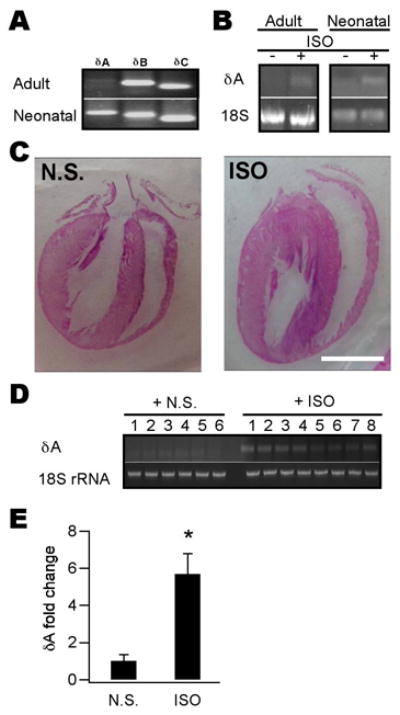
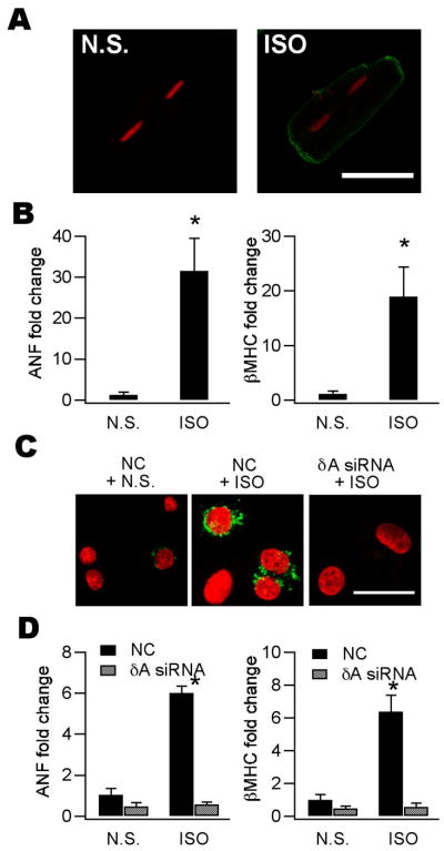
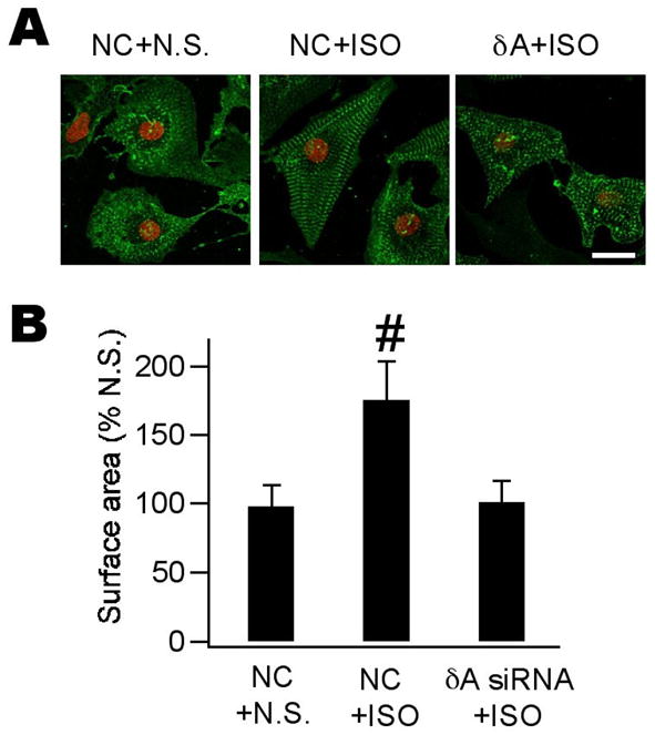
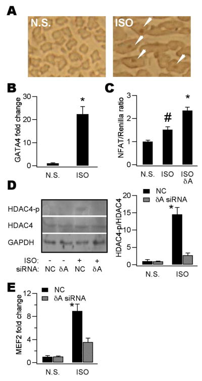
Ca(2+)/calmodulin-dependent protein kinase II (CaMKII) is a new promising target for prevention and treatment of cardiac hypertrophy and heart failure. There are three δ isoforms of CaMKII in the heart and previous studies focused primarily on δB and δC types. Here we report the δA isoform of CaMKII is also critically involved in cardiac hypertrophy. We found that δA was significantly upregulated in pathological cardiac hypertrophy in both neonatal and adult models. Upregulation of δA was accompanied by cell enlargement, sarcomere reorganization and reactivation of various hypertrophic cardiac genes including atrial natriuretic factor (ANF) and β-myocin heavy chain (β-MHC). Studies further indicated the pathological changes were largely blunted by silencing the δA gene and an underlying mechanism indicated selective interference with the HDAC4-MEF2 signaling pathway. These results provide new evidence for selective interfering cardiac hypertrophy and heart failure when CaMKII is considered as a therapeutic target.


Figures

Figure 1
Figure 1. The δA isoform was upregulated in various cardiac hypertrophy models
A, mRNAs of 3 CaMKII δ-isoforms were detected using RT-PCR in isolated adult (upper) and cultured neonatal (lower) myocytes. δA was not detectable in adult cells. B, CaMKIIδA mRNA in adult and neonatal myocytes was upregulated by ISO (10 μM) treatment for 24 h. 18-S rRNA was used as an internal control. C, Representative four-chambered heart pictures showing typical cardiac hypertrophy induced by 5-d injection of ISO (30 mg kg−1 d−1, i.p.). The hearts were sectioned and stained by the H&E method. Scale bar = 1 cm. D, CaMKIIδA mRNA (upper) from ISO-injected animal’s heart tissue (n = 8) was upregulated compared to that of NS-injected ones (n = 6). 18-S rRNA was used as a loading control (lower). E, Quantitative analysis of data shown in (D). Band density of CaMKIIδA for each sample was determined using ImageJ and normalized to their respective 18-S rRNA values. (n = 6–8, *p < 0.01, Student’s t test).
Figure 2
Figure 2. Reactivation of fetal cardiac genes was blunted by CaMKIIδA silencing
A, induction and immunolocalization of ANF protein in isolated adult myocytes treated by ISO (2 μM) for 48 h. For immunofluorescence cells were first incubated with an anti-ANF antibody followed by reaction with Alexa-488 conjugated second antibody (green). Propidium iodine (PI) was used as counterstain for nuclei (red). Scale bar = 50 μm. B, Upregulation of ANF and β–MHC mRNAs in the hearts from ISO-injected animals. mRNAs were analyzed by qPCR and normalized to 18-S rRNA. The mean normalized value for expression of each gene in NS-injected animals is defined as 1. (n = 3–4, *p < 0.01, Student’s t-test). C, induction and immunolocalization of ANF protein in cultured neonatal myocytes by ISO (10 μM) for 48 h. Note the ANF signal is primarily located in the perinuclear area. Scale bar = 20 μm. D. qPCR assays showing both ANF and βMHC mRNAs were upregulated by ISO (10 μM) application for 48 h and these effects were blunted by δA siRNA. (for both panels n = 3, *p < 0.01 vs NC/NS group, ANOVA).
Figure 3
Figure 3. Morphological alterations were prevented by CaMKIIδA silencing
A, confocal images of neonatal myocytes with various treatments as indicated. Cells were treated with N.S. or ISO for 48 h and incubated with anti-α-actinin followed by reaction with Alexa-488 conjugated second antibody (green). Propidium iodine was used as counterstain for nuclei (red). Scale bar = 50 μm. ISO induced cell enlargement accompanied by prominent sarcomere reorganization (middle). These alterations are largely prevented by pretreatment with δA siRNA (right). B, quantitative analysis of data shown in (A). At least 100 cells were quantified for each group and experiments were repeated twice. (n = 3, #p < 0.05, ANOVA).
Figure 4
Figure 4. CaMKIIdA silencing blunted activation of HDAC4-MEF2 signaling
A, in situ detection of nuclear NFAT protein (arrows in right panel) using immunohistochemistry in ISO-induced hypertrophy models. In NS-injected animals, no appreciable NFAT signal was revealed (left). Note the larger cell size in right panel. B, GATA-4 mRNA was significantly amplified in heart tissues of ISO-treated animals (n = 6, *p < 0.01, Student’s t test). C, NFAT-mediated transcription activity as reflected by the NFAT-luciferase assay was increased by ISO alone and further enhanced by CaMKIIδA silencing. (n = 3, #p < 0.05 vs N.S.; *p < 0.05 vs ISO alone, ANOVA). These experiments and what follows were performed in neonatal myocytes. D, Western blot showing profound phosphorylation of HDAC4 by 48-h ISO incubation. CaMKIIδA silencing prevented HDAC4 phosphorylation evoked by ISO (left). Plotted on the right is quantitative analysis of data shown in the left panel. Band density was determined and analyzed using ImageJ. (n = 4, *p < 0.01 vs NC/NS group, ANOVA). E, MEF2 mRNA was upregulated by ISO incubation for 48 h and this effect was blunted by CaMKIIδA silencing. (n = 4, *p < 0.01 vs NC/NS group, ANOVA).

Similar articles

Cited by

References

    1. Sambrano GR, Fraser I, Han H, Ni Y, O’Connell T, Yan Z, Stull JT. Navigating the signalling network in mouse cardiac myocytes. Nature. 2002;420:712–714. - PubMed
    1. Frey N, Olson EN. Cardiac hypertrophy: the good, the bad, and the ugly. Annu Rev Physiol. 2003;65:45–79. - PubMed
    1. Bers DM. Altered cardiac myocyte Ca2+ regulation in heart failure. Physiology (Bethesda) 2006;21:380–387. - PubMed
    1. Anderson ME. CaMKII and a failing strategy for growth in heart. J Clin Invest. 2009;119:1082–1085. - PMC - PubMed
    1. Bossuyt J, Helmstadter K, Wu X, Clements-Jewery H, Haworth RS, Avkiran M, Martin JL, Pogwizd SM, Bers DM. Ca2+/calmodulin-dependent protein kinase IIδ and protein kinase D overexpression reinforce the histone deacetylase 5 redistribution in heart failure. Circ Res. 2008;102:695–702. - PubMed

Publication types

MeSH terms

LinkOut - more resources