Review
. 2011 Sep;131(3):269-86.
doi: 10.1016/j.pharmthera.2011.04.008. Epub 2011 Apr 28.

Promise of resveratrol for easing status epilepticus and epilepsy

Affiliations
Review

Promise of resveratrol for easing status epilepticus and epilepsy

Ashok K Shetty. Pharmacol Ther. 2011 Sep.

Abstract

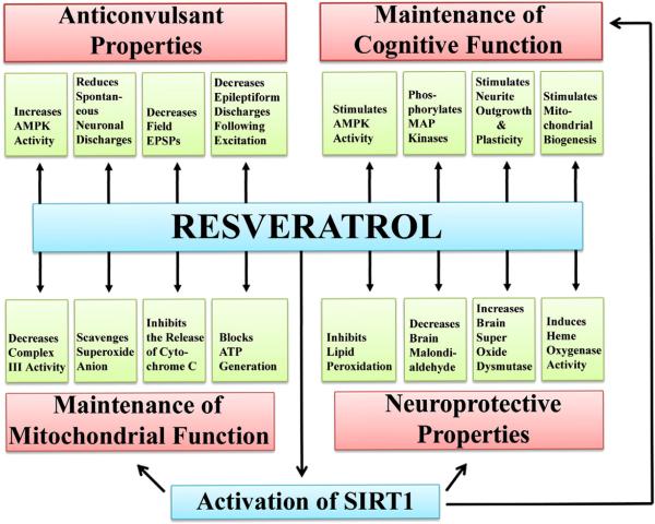
Resveratrol (RESV; 3,5,4'-tri-hydroxy stilbene), a naturally occurring phytoalexin, is found at a high concentration in the skin of red grapes and red wine. RESV mediates a wide-range of biological activities, which comprise an increased life span, anti-ischemic, anti-cancer, antiviral, anti-aging and anti-inflammatory properties. Studies in several animal prototypes of brain injury suggest that RESV is an effective neuroprotective compound. Ability to enter the brain after a peripheral administration and no adverse effects on the brain or body are other features that are appealing for using this compound as a therapy for brain injury or neurodegenerative diseases. The goal of this review is to discuss the promise of RESV for treating acute seizures, preventing the acute seizure or status epilepticus induced development of chronic epilepsy, and easing the chronic epilepsy typified by spontaneous recurrent seizures and cognitive dysfunction. First, the various beneficial effects of RESV on the normal brain are discussed to provide a rationale for considering RESV treatment in the management of acute seizures and epilepsy. Next, the detrimental effects of acute seizures or status epilepticus on the hippocampus and the implications of post-status epilepticus changes in the hippocampus towards the occurrence of chronic epilepsy and cognitive dysfunction are summarized. The final segment evaluates studies that have used RESV as a neuroprotective compound against seizures, and proposes studies that are critically needed prior to the clinical application of RESV as a prophylaxis against the development of chronic epilepsy and cognitive dysfunction after an episode of status epilepticus or head injury.


Figures

Fig. 1
Fig. 1
The various beneficial effects of resveratrol (RESV) on the normal brain function that provide a strong rationale for administering RESV in conditions such as status epilepticus (SE) or brain injury as a neuroprotective compound. The neuroprotective properties of RESV are supported by its ability to inhibit the lipid peroxidation, reduce brain malondialdehyde levels, enhance the concentration of brain superoxide dismutase, and induce heme oxygenase activity (see the lower right region of the illustration). The role of RESV in the maintenance of mitochondrial function is indicated by its ability to decrease complex III activity, scavenge superoxide anions, inhibit the release of cytochrome C, and block adenosine triphosphate (ATP) generation (see the lower left region). The anticonvulsant properties of RESV are typified by its ability to increase 5′ AMP-activated protein kinase (AMPK) activity, reduce spontaneous neuronal discharges, decrease the field excitatory post-synaptic potentials, and decrease the epileptiform discharges following excitation (see the upper left region). The contribution of RESV towards the maintenance of cognitive function is supported by its ability to stimulate AMPK activity, phosphorylate mitogen-activated protein (MAP) kinases, stimulate the neurite outgrowth, and promote neural plasticity and mitochondrial biogenesis (see the upper right region).
Fig. 2
Fig. 2
The structure of the hippocampus at 4 days after status epilepticus (SE) when visualized with the neuron-specific nuclear antigen (NeuN) immunostaining. A1 and A3 show anterior and posterior regions of the hippocampus from a vehicle-treated control rat. B1 and B3 show anterior and posterior regions of the hippocampus from a KA-treated rat showing moderate hippocampal injury. C1 and C3 show hippocampal regions from a KA treated rat showing massive hippocampal injury. A2, B2 and C2 are magnified views of dentate hilar (DH) regions from A1, B1 and C1. Note that, in the moderate injury group (B1B3), the loss of neurons is considerable in the dentate hilus (DH) and the CA1 subfield but modest in the CA3 region. In contrast, in the massive injury group (C1C3), the loss of CA1 and CA3 pyramidal neurons is dramatic throughout the hippocampus. DG, dentate gyrus. Scale bar, A1, B1, C1 and A3, B3 and C3=500 μm; A2, B2 and C2=100 μm. The bar chart (D) depicts the absolute number of surviving neurons in different regions of the hippocampus of vehicle-treated rats, and KA-treated rats with moderate or massive hippocampal injury at 4 days post-administration. The loss of neurons in KA treated rats with moderate hippocampal injury is significant in the dentate hilus, and CA1 and CA3 pyramidal cell layers. However, there is no loss of neurons in the granule cell layer. The massive injury group exhibits greater loss of CA1 and CA3 pyramidal neurons than the moderate injury group. Moreover, the dentate gyrus of the massive injury group exhibits significant loss of both dentate granule cells and dentate hilar neurons. DH, dentate hilus; GCL, granule cell layer.
Fig. 3
Fig. 3
The extent of inflammation in the hippocampus after an episode of status epilepticus (SE) when visualized with the immunostaining for ED-1 antigen (a trans-membrane protein that identifies activated microglial cells). Figure A1 illustrates a higher density of activated microglia in regions of the neurodegeneration such as the dentate hilus (DH) and the CA1 subfield. A2 is magnified view of activated microglia from a region of the DH. For ED-1 immunostaining methods, see Hattiangady et al. (2011). Scale bar, A1=500 μm; A2=100 μm.
Fig. 4
Fig. 4
Status epilepticus impairs the ability for spatial learning and spatial memory retrieval even at extended time-points after an episode of SE. The figure A on the left side compares the spatial learning ability of rats that underwent SE and developed chronic epilepsy at ~4-months after SE (indicated by a red line) with the age-matched naive rats (indicated by a green line) in a water maze test (WMT). Note that, in comparison to intact rats, the average swim path lengths to reach the platform were much greater in rats exhibiting chronic epilepsy in all of the eleven training sessions (A). Naïve rats learn quickly to locate the hidden platform using spatial cues and hence their swim path lengths are much shorter after 3–4 sessions of learning. The epileptic rats are clearly slow learners and exhibit overnight forgetting. The figures in B and C show the results of a probe test (i.e. a 30 second memory retrieval test for each rat without the hidden platform) conducted one-day after the eleven learning sessions performed over 6 days. The circular diagram on the left side of figure B depicts the various water maze tank quadrants in different colors and the swim path of a naïve rat. Note that this naïve rat exhibits robust memory retention, as it swam straight to the quadrant where the platform was originally placed (gray-colored area) and spent most of its probe test time searching for the platform in this quadrant. The circular diagram on the right side of figure B illustrates the swim path of a chronically epileptic rat in various quadrants of the water maze tank. Note that this epileptic rat explored all quadrants of the maze without any specific affinity for the quadrant where the platform was originally placed. The bar chart in C compares the dwell time of rats from the naïve group and the epileptic group in different quadrants of the water maze tank. Note that control rats spend most of their probe test time in the platform quadrant (gray colored bar) whereas epileptic rats spend more or less equal amount of time in all four quadrants, clearly implying that chronically epileptic rats exhibit memory dysfunction. For methods pertaining to water maze tests, see Hattiangady et al. (2011).
Fig. 5
Fig. 5
Changes in the dentate neurogenesis after an episode of status epilepticus (SE) induced by kainic acid. Newly born neurons in the dentate gyrus (DG) of a naïve adult rat (A1) and an adult rat that underwent status epilepticus 12 days prior to euthanasia (B1) were visualized with immunostaining for doublecortin (a marker of newly born neurons). A2 and B2 show magnified views of regions of dentate gyrus from A1 and B1 respectively. Note that, in comparison to the dentate gyrus of a control rat (A1, A2), a rat that underwent SE (B1, B2) exhibits considerably increased density of doublecortin + new neurons and abnormal migration of newly born neurons into the dentate hilus (indicated by arrowheads in B1). C1 is magnified view of a region from B1 showing aberrantly migrated newly born neurons in the dentate hilus. DH, dentate hilus; GCL, granule cell layer; SGZ, subgranular zone. Scale bar, A1 and B1=200 μm; A2, B2 and C1=50 μm.
Fig. 6
Fig. 6
Status of dentate neurogenesis in chronic epilepsy as revealed by doublecortin (DCX) immunostaining. A1–C1 illustrate the distribution of DCX positive newly born neurons in the dentate gyrus of an age-matched intact rat (A1), a rat exhibiting chronic epilepsy at 5 months after an intracerebroventricular (ICV) kainic acid (KA) administration (B1), and a rat displaying robust chronic epilepsy at 5-months after intraperitoneal (IP) KA-induced status epilepticus (SE) (C1). Note that, in comparison to the age-matched intact hippocampus, hippocampi from chronically epileptic animals exhibit dramatically reduced density of DCX positive newly generated neurons. Arrowheads point to regions in the subgranular zone (SGZ) where neurogenesis is active. The arrow in C1 denotes a neuron that has migrated into the dentate hilus. A2, B2, and C2 are magnified views of regions from A1, B1, and C1 demonstrating the morphology newly generated neurons in the three groups. In the dentate gyrus of the age-matched intact rat (A2), DCX positive new neurons exhibit long apical dendrites that extend into the molecular layer (ML) through the granule cell layer (GCL). Contrastingly, in hippocampi from epileptic animals (B2, C2), a vast majority of DCX positive neurons display basal dendrites (indicated by short arrows). The bar chart compares the absolute numbers of DCX positive new neurons in different groups. Note that, in comparison to the age-matched control rats, the overall dentate neurogenesis in hippocampi of chronically epileptic rats is drastically reduced. Furthermore, the decline is more pronounced in the hippocampus of rats exhibiting robust chronic epilepsy (i.e. the hippocampus at 5-months post-SE) than the hippocampus of rats exhibiting fewer spontaneous seizures (i.e. the hippocampus at 5-months post-ICV KA administration). DH, Dentate hilus; GCL, Scale bar, A1, B1, C1=200 μm; A2, B2, C2=50 μm.
Fig. 7
Fig. 7
Differentiation of newly born cells into neurons, astrocytes, and oligodendrocyte progenitors at 24 h after twelve daily injections of BrdU in the chronically epileptic hippocampus. Examples of newly born cells that differentiate into doublecortin + (DCX+) immature neuron (A1–A3), TuJ-1+ neuron (B1; indicated by an arrow), S-100β+ astrocyte (C1; denoted by an arrow), and NG2+ oligodendrocyte progenitor (D1; showed by an arrow) in the subgranular zone-granule cell layer (SGZ-GCL) are illustrated. Scale bar, A1–A3=5 μm; B1, C1, D1=10 μm. The bar chart in E1 compares percentages of newly born cells (i.e. BrdU + cells) that express DCX, TuJ-1, S-100β or NG2 in the subgranular zone-granule cell layer (SGZ-GCL) between the age-matched intact hippocampus and the chronically epileptic hippocampus. Note that the neuronal differentiation of newly born cells is dramatically decreased but differentiation of newly added cells into S-100β+ or NG2+ glia is considerably enhanced in the chronically epileptic hippocampus.
Fig. 8
Fig. 8
The upper panel shows that, status epilepticus (SE) depletes the population of GABA-ergic interneurons expressing the neuropeptide, the neuropeptide Y (NPY). Figure A illustrates the distribution and density of NPY immunopositive interneurons in the dentate hilus of a naïve rat. Figure B shows the distribution and density of NPY + interneurons in the dentate hilus of an age-matched rat that underwent kainic acid induced SE and acquired chronic epilepsy. Note that the overall density of NPY + interneurons is greatly reduced after SE. For NPY immunostaining methods see Rao et al. (2006). Scale bar, A1 and A2=200 μm. The lower panel illustrates the extent of the aberrant mossy fiber sprouting in rats with moderate hippocampal injury (B1–B2) and rats with severe hippocampal injury (C1–C2), in comparison to age-matched intact rats (A1–A2), visualized by Timm's histochemical staining. Note that, in comparison to rats exhibiting moderate hippocampal injury (B1–B2), the rats showing severe hippocampal injury (C1–C2) have much robust aberrant sprouting of mossy fibers into the dentate supragranular layer (DSGL). Asterisks in C1 denote mossy fiber sprouting in the DSGL. DH, dentate hilus; GCL, granule cell layer. Scale bar, A1, B1 & C1=500 μm; A2, B2 and C2=200 μm.
Fig. 9
Fig. 9
Electrical activity in the hippocampus during a spontaneous seizure in a chronically epileptic rat, as recorded through electroencephalography (EEG). The activity was recorded through an electrode implanted into the dentate gyrus. Note the presence of persistent large amplitude and high frequency polyspikes for over 50 s. Polyspikes represent a complex paroxysmal EEG pattern with close association of two or more diphasic spikes occurring more or less rhythmically in bursts of variable duration, generally with large amplitudes. For methods of electrode implantation into the dentate gyrus, see Rao et al. (2006).
Fig. 10
Fig. 10
Potential outcome of resveratrol (RESV) therapy for status epilepticus (SE) and epilepsy. The overall outcome of RESV treatment would likely depend on the time-point of its administration after an initial precipitating injury (IPI) such as SE or head injury. An IPI such as SE leads first to changes that comprise increased excitation of neurons, enhanced oxidative stress, considerable neurodegeneration, inflammatory reaction and abnormal neurogenesis, which are broadly classified as “early changes”. While most of these changes can occur in several brain regions, the hippocampus has been recognized as the most vulnerable region to SE for exhibiting all of these changes. This initial phase is typically followed by an intermediate “epileptogenesis” phase during which the various epileptogenic changes occur. These include the loss of GABA-ergic interneurons, decreases in the functional inhibition, aberrant sprouting of axons, and abnormal integration of newly born neurons. All of these changes are believed to contribute towards the development of an epileptogenic circuitry, which gradually leads to a state of “chronic epilepsy” typified by spontaneous recurrent seizures, decreased hippocampal neurogenesis and cognitive dysfunction. Considering the above, the commencement of RESV treatment immediately after the induction of SE may either prevent or greatly minimize the early changes, which may block “epileptogenesis” as well as the evolution of SE into “chronic epilepsy”. This hypothesis is based on the anticonvulsant, anti-oxidant, anti-inflammatory, and anti-apoptotic properties of RESV. On the other hand, the commencement of RESV treatment after “early changes” may reduce the extent of “epileptogenesis” through preservation of greater numbers of GABA-ergic interneurons, dampening of hippocampal hyperexcitability and maintenance of normal neurogenesis. These actions of RESV may considerably restrain the development/intensity of chronic epilepsy and cognitive dysfunction. This premise is based on the neuroprotective, anti-inflammatory and anticonvulsant properties of RESV. Furthermore, the commencement of RESV treatment after most of the epileptogenic changes occurs or after the establishment of a chronic epileptic state may still restrain the intensity of chronic epilepsy and improve the cognitive function. This proposition is based on the anti-inflammatory and anticonvulsant properties of RESV and the ability of RESV to activate SIRT1.

Similar articles

Cited by

References

    1. Abd El-Mohsen M, Bayele H, Kuhnle G, Gibson G, Debnam E, Kaila Srai S, et al. Distribution of [3 H]trans-resveratrol in rat tissues following oral administration. Br J Nutr. 2006;96:62–70. - PubMed
    1. Abrous DN, Koehl M, Le Moal M. Adult neurogenesis: From precursors to network and physiology. Physiol Rev. 2005;85:523–569. - PubMed
    1. Aimone JB, Wiles J, Gage FH. Potential role for adult neurogenesis in the encoding of time in new memories. Nat Neurosci. 2006;9:723–727. - PubMed
    1. Alessio A, Damasceno BP, Camargo CH, Kobayashi E, Guerreiro CA, Cendes F. Differences in memory performance and other clinical characteristics in patients with mesial temporal lobe epilepsy with and without hippocampal atrophy. Epilepsy Behav. 2004a;5:22–27. - PubMed
    1. Alessio A, Kobayashi E, Damasceno BP, Lopes-Cendes I, Cendes F. Evidence of memory impairment in asymptomatic individuals with hippocampal atrophy. Epilepsy Behav. 2004b;5:981–987. - PubMed

Publication types