. 2011 Aug;278(1-2):77-85.
doi: 10.1016/j.heares.2011.04.011. Epub 2011 May 1.

Activity of all JNK isoforms contributes to neurite growth in spiral ganglion neurons

Affiliations

Activity of all JNK isoforms contributes to neurite growth in spiral ganglion neurons

Patrick J Atkinson et al. Hear Res. 2011 Aug.

Abstract

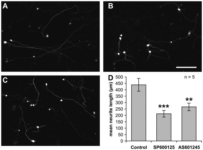
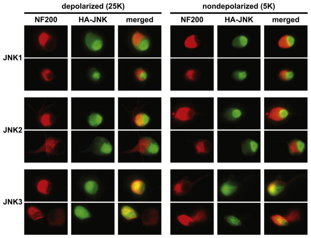
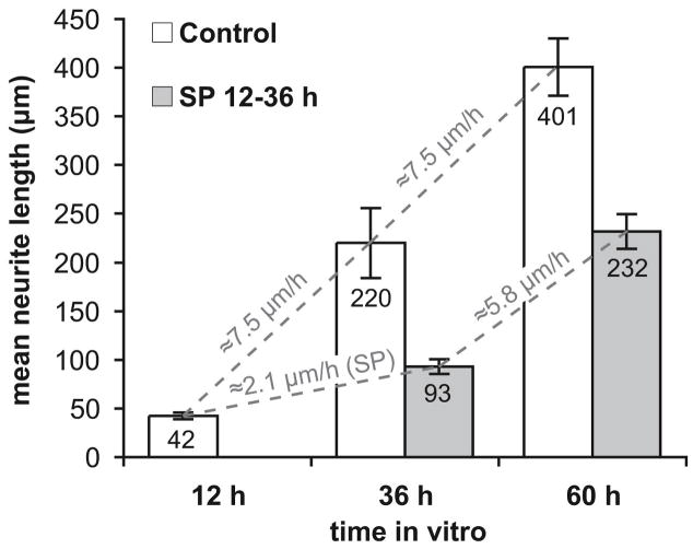
Jun N-terminal kinase (JNK) is a multifunctional protein kinase crucial for neuronal apoptosis as well as neurite growth. We have previously shown that JNK activity is correlated with spiral ganglion neuron (SGN) apoptosis following hair cell loss in rats (Alam et al., 2007) implying that JNK inhibition may have therapeutic potential to protect SGNs in deaf individuals. Here we investigated the role of JNK in neurite outgrowth from cultured neonatal rat and mouse SGNs. We show that JNK is required for initial growth of neurites and for continued extension of already established neurites. The effect of JNK inhibition on neurite growth is rapid and is also rapidly reversible after washout of the inhibitor. Using phosphoJNK immunoreactivity as an indicator, we show that JNK is activated in growth cones within 30 min after transfer to medium lacking neurotrophic stimuli (5 K medium) but activation in the nucleus and soma requires hours. By transfecting epitope-tagged JNK1, JNK2, or JNK3 isoforms into SGNs, we found that all are present in the nucleus and cytoplasm and that there is no preferential redistribution to the nucleus after transfer to 5 K medium. Cotransfection of dominant-negative (dn) JNK1 and JNK2 into SGNs reduced neurite growth, although transfection of dnJNK1 or dnJNK2 alone had no significant effect. SGNs cultured from JNK3(-/-) mice showed reduced neurite growth that was further reduced by transfection of dnJNK1 and dnJNK2. This indicates that all three JNK isoforms promote SGN neurite growth although there may be functional redundancy between JNK1 and JNK2.


Figures

Figure 1
Figure 1
JNK inhibitor, SP600125 or AS601245 (both at 20 μM) were added to spiral ganglion cultures after 24 h in vitro. The cultures were fixed after an additional 24 h, labeled with ant-NF200 and imaged. A–C are representative low-magnification (scale bar = 150 μm) images of SGNs cultured in control conditions (A), SP600125 (B), or AS601245 (C). D: quantitation of neurite lengths. Neurite growth was significantly (**p<0.01, ***p<0.001) decreased after the addition of the either JNK inhibitor. Shown are means ± SEM (n = 5 replicate cultures).
Figure 2
Figure 2
Spiral ganglion cultures were fixed 36 h or 60 h after plating. SP600125 (20 μM) was present for 24 h – either from 12 h to 36 h in culture or from 36 h to 60 h – and the cultures were fixed immediately after for comparison to control cultures fixed at the same time but not treated with SP600125. Shown are mean neurite lengths ± SEM (n = 5 replicate cultures). JNK inhibition significantly (**p<0.01) inhibited neurite growth whether present 12–36 h after plating, when neurite growth is beginning, or 36–60 hr after plating, when neurites are already established. The approximate mean rate of neurite growth is shown in gray lettering for 24 h time intervals in which SP600125 was either present or absent.
Figure 3
Figure 3. Representative images of HA-tagged JNK fusion proteins in cultured SGNs
Cells were maintained for 24 h in depolarizing (25K) conditions for trophic support and then incubated for an additional 6 h in 25K or without trophic support in nondepolarizing serum-free medium, as indicated. Cultures were then fixed and neurons visualized with anti-NF200 (red) and HA-JNKs with anti-HA (green). Two typical examples are shown for each isoform. The bottom row shows examples of two adjacent neurons of which one was transfected and one not. All JNK isoforms are distributed throughout the cytoplasm and nucleus, although JNK1 and JNK2 are more strongly localized to the nucleus than is JNK3. The subcellular distribution of all JNKs is not apparently affected by withdrawal of trophic support. Scale: individual images are 30 μm × 40 μm.
Figure 4
Figure 4. Representative images of JNK phosphorylation in SGN somata and growth cones
A–F: JNK is phosphorylated in the soma and nucleus of a SGN in vitro, but not in adjacent glia, six hours after transfer to medium lacking neurotrophic support (5K, A–C) from depolarizing (25K, D–F) medium. Scale bar = 10 μm. A,D: NF200 (green) and bisbenzamide nuclear label (blue). B,E: pJNK immunofluorescence (red). C,F: merged NF200, nuclear and pJNK labeling. G–L: JNK is phosphorylated in a punctate pattern in the growth cone 30 minutes after transfer to medium lacking neurotrophic support (5K, G–I) from depolarizing (25K, J–L) medium. Scale bar = 5 μm. G,J: Lck-GFP (green). H,K: pJNK immunofluorescence (red). I,L: merged Lck-GFP and pJNK labeling.
Figure 5
Figure 5
JNK phosphorylation was assayed by measuring phosphoJNK immunofluorescence (IF) in growth cones (A) or somata (B,C), at 30 min (A,B) or 6 h (C) after shift from 25K to one of the following experimental conditions: 25K control; 5K DMEM lacking neurotrophic factors; 5K with 100 ng/ml NT-3; 5K with 20 μM SP600125 (SP). The data were normalized to phosphoJNK IF in the 5K condition in each experiment. JNK phosphorylation is significantly elevated in growth cones within 30 min of trophic factor deprivation by shift to 5K but the increase is prevented by NT-3 or by SP600125. In contrast, JNK phosphorylation is not elevated in somata 30 min after shift to 5K but is elevated 6 h after shift. Statistical significance of differences relative to 5K was determined by ANOVA with Tukey-Kramer correction (*p<0.05, **p<0.01, ***p<0.001).
Figure 6
Figure 6. Inhibition of SGN neurite growth by JNK inhibitor is reversible
Cultures were fixed 12, 36 or 60 hours after plating. Control cultures were maintained in 25K/0.1% DMSO. SP600125-treated cultures had SP600125 present from 12 to 36 h after plating. Shown are means ± SEM (n = 5 replicate cultures) with the mean neurite length printed in each bar. The approximate mean rate of neurite growth is shown in gray for each 24 h interval, 1236 h and 3660 h post-plating. This was determined by dividing the difference in mean neurite length by 24 h. SP600125 reduces the neurite growth rate when present in the 12–36 h interval but growth returns to a nearly control rate in the 36–60 h during which SP600125-treated SGNs had 24 h to extend neurites after SP600125 withdrawal.
Figure 7
Figure 7
A–E: SGNs were cultured either from JNK3+/+ (WT) or JNK3−/− (KO) littermate mice and transfected with either eGFP and empty vector plasmid (1:4 ratio of eGFP:vector) or with eGFP and dnJNK1 and dnJNK2 (1:2:2 ratio). Cotransfection of eGFP identified transfected cells. These combinations resulting in neurons expressing either all three JNK isoforms (JNK1,2,3), or only JNK1 and JNK2 (JNK1,2), or only JNK3, or no JNK (none). A–D: Representative images of cultured mouse SGNs labeled by eGFP fluorescence and transfected with vector (A,B) or dnJNK1 and dnJNK2 (C,D). A and C are from WT mice, B and D are from JNK3 KO mice. Scale bars = 50 μm. E. Neurite growth over a 24 h period was quantified. There is a significant increasing decrement in neurite growth with removal of additional JNK isoforms. F. SGNs were cultured from neonatal rats and transfected with eGFP and empty vector, dnJNK1, dnJNK2, dnJNK1 and dnJNK2 combined, or MKK-JNK1. Neurite lengths were determined after 48 h. There was no significant difference in mean neurite length between vector and a single dnJNK (horizontal bar). Combined dnJNK1 and dnJNK2 significantly reduced neurite length and constitutively-active JNK (MKK-JNK1) significantly increased neurite length. For E and F, error bar shows SEM. The number of independent replicate cultures is given within each bar and statistical significance was determined by ANOVA with a Tukey-Kramer post-hoc test, *p<0.05, **p<0.01, ***p<0.001.

Similar articles

Cited by

References

    1. Alam SA, Robinson BK, Huang J, Green SH. Prosurvival and proapoptotic intracellular signaling in rat spiral ganglion neurons in vivo after the loss of hair cells. J Comp Neurol. 2007;503:832–852. - PubMed
    1. Amagasaki K, Kaneto H, Heldin CH, Lennartsson J. c-Jun N-terminal kinase is necessary for platelet-derived growth factor-mediated chemotaxis in primary fibroblasts. J Biol Chem. 2006;281:22173–9. - PubMed
    1. Barnat M, Enslen H, Propst F, Davis RJ, Soares S, Nothias F. Distinct roles of c-Jun N-terminal kinase isoforms in neurite initiation and elongation during axonal regeneration. J Neurosci. 2010;30:7804–16. - PMC - PubMed
    1. Benediktsson AM, Schachtele SJ, Green SH, Dailey ME. Ballistic labeling and dynamic imaging of astrocytes in organotypic hippocampal slice cultures. J Neurosci Methods. 2005;141:41–53. - PubMed
    1. Bennett BL, Sasaki DT, Murray BW, O’Leary EC, Sakata ST, Xu W, Leisten JC, Motiwala A, Pierce S, Satoh Y, Bhagwat SS, Manning AM, Anderson DW. SP600125, an anthrapyrazolone inhibitor of Jun N-terminal kinase. Proc Natl Acad Sci U S A. 2001;98:13681–6. - PMC - PubMed

Publication types

MeSH terms

Substances