. 2011 Jul 1;51(1):179-84.
doi: 10.1016/j.freeradbiomed.2011.04.004. Epub 2011 Apr 15.

Febuxostat inhibition of endothelial-bound XO: implications for targeting vascular ROS production

Affiliations

Febuxostat inhibition of endothelial-bound XO: implications for targeting vascular ROS production

Umair Z Malik et al. Free Radic Biol Med. .

Abstract

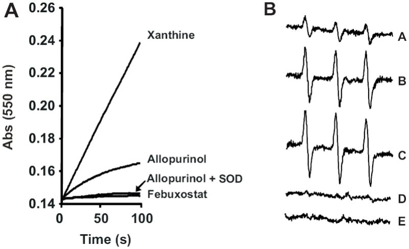
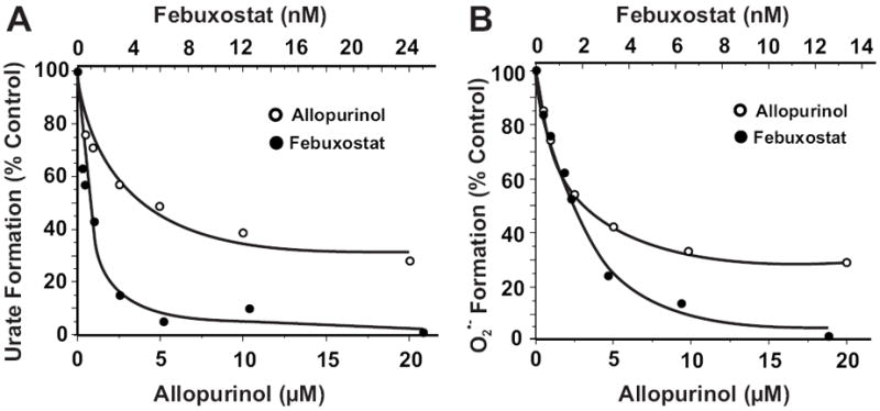
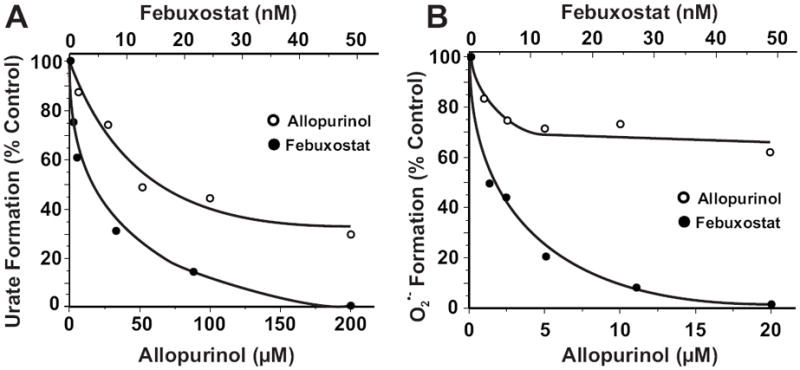
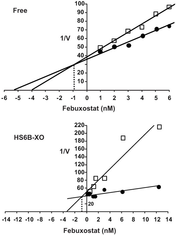
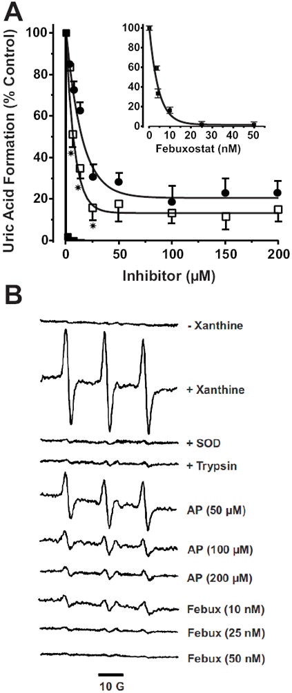
Xanthine oxidase (XO) is a critical source of reactive oxygen species (ROS) that contribute to vascular inflammation. Binding of XO to vascular endothelial cell glycosaminoglycans (GAGs) results in significant resistance to inhibition by traditional pyrazolopyrimidine-based inhibitors such as allopurinol. Therefore, we compared the extent of XO inhibition (free and GAG-bound) by allopurinol to that by febuxostat, a newly approved nonpurine XO-specific inhibitor. In solution, febuxostat was 1000-fold more potent than allopurinol at inhibiting XO-dependent uric acid formation (IC₅₀= 1.8 nM vs 2.9 μM). Association of XO with heparin-Sepharose 6B (HS6B-XO) had minimal effect on the inhibition of uric acid formation by febuxostat (IC₅₀= 4.4 nM) while further limiting the effect of allopurinol (IC₅₀= 64 μM). Kinetic analysis of febuxostat inhibition revealed K(i) values of 0.96 (free) and 0.92 nM (HS6B-XO), confirming equivalent inhibition for both free and GAG-immobilized enzyme. When XO was bound to endothelial cell GAGs, complete enzyme inhibition was observed with 25 nM febuxostat, whereas no more than 80% inhibition was seen with either allopurinol or oxypurinol, even at concentrations above those tolerated clinically. The superior potency for inhibition of endothelium-associated XO is predictive of a significant role for febuxostat in investigating pathological states in which XO-derived ROS are contributive and traditional XO inhibitors are only slightly effective.


Figures

Fig. 1
Fig. 1. Reaction of allopurinol with XO induces ROS formation
(A) XO (10 mU/ml) was exposed to either xanthine (50 μM), allopurinol (50 μM), Febuxostat (100 nM) or allopurinol (50 μM) + SOD (10 U/ml) and O2 •- formation monitored by the reduction of cytochrome c (λ = 550 nm) over time. (B) Xanthine oxidase (10 mU/ml) was exposed to allopurinol (50 μM) and O2 •- formation monitored by EPR spin trapping with PPH (100 μM). Spectra represent the following conditions: allopurinol 25, 50 and 100 s (A-C, respectively), 100 s + SOD (D) and 100 s + febuxostat (E).
Fig. 2
Fig. 2. Febuxostat is more potent than allopurinol at inhibiting XO free in solution
(A) Xanthine oxidase (2 mU/ml, 5 mM KPi, pH 7.4) was exposed to various concentrations of either allopurinol or febuxostat and assessed for formation uric acid (λ = 295 nm) upon the addition of xanthine (50 μM). Shown are the initial reaction rates (V0) plotted as % control (no inhibitor). IC50 values were calculated as the ordinate value of 50% inhibition (allopurinol = 2.9 μM and febuxostat = 1.8 nM). (B) Same as A except O2 •- formation was detected by the reduction of cytochrome c (λ = 550) and IC50 values were 3.9 μM for allopurinol and 0.9 nM for febuxostat.
Fig. 3
Fig. 3. Febuxostat inhibition of GAG-immobilized XO is superior to allopurinol
(A) HS6B-XO (2 mU/ml, 5 mM KPi, pH 7.4) was exposed to various concentrations of either allopurinol or febuxostat and assessed for formation of uric acid (λ = 295 nm) upon the addition of xanthine (200 μM). Shown are the initial reaction rates (V0) plotted as % control (no inhibitor). IC50 values were calculated as the ordinate value of 50% inhibition (allopurinol = 64 μM and febuxostat = 4.4 nM). (B) Same as A except O2 •- formation was detected by the reduction of cytochrome c (λ = 550 nm) and IC50 values were 77 μM for allopurinol and 4.6 nM for febuxostat.
Fig. 4
Fig. 4. Kinetic analysis of XO inhibition by febuxostat
(A) Xanthine oxidase (2 mU/ml, 5 mM KPi, pH 7.4) was exposed to various concentrations of febuxostat and assessed for formation uric acid (λ = 295) using 2 concentrations of xanthine (10 and 100 μM). Shown is a Dixon plot (1/V vs. [inhibitor]) generating an inhibition constant (Ki) = 0.96 nM for febuxostat. (B) HS6B-XO (2 mU/ml, 5 mM KPi, pH 7.4) was used with xanthine (10 and 100 μM) generating an inhibition constant (Ki) = 0.92 nM for febuxostat. The Ki values were calculated as the ordinate value of the intersection of the 2 lines.
Fig. 5
Fig. 5. Febuxostat is superior to allo/oxypurinol at inhibiting endothelial cell-bound XO
A) BAECs were exposed to purified XO (5 mU/ml) for 20 min 25°C and washed as described in the methods. Inhibitors were added at the indicated concentrations followed by xanthine (100 μM) and uric acid levels assessed after 1 h. Values represent % control (no inhibitor) and are the mean of at least 3 independent determinations (* = p < 0.05). B) Purified XO was bound to BAEC GAGs (as in A), the cells were then harvested by mechanical dissociation and resuspended at 1 × 10⁶ cells/ml. Cell suspensions were exposed to the indicated concentrations of inhibitor followed by xanthine (100 μM) and immediately analyzed by EPR spin trapping of extracellular O2 •- with PPH (50 μM). Spectra represent XO-loaded cells exposed to PPH and the following agents from top to bottom: (- xanthine), (+ xanthine), (+ xanthine and SOD), (100 U/ml), (XO-loaded cells treated with trypsin to remove extracellular XO and then exposed to xanthine), (+ xanthine and 50, 100 or 200 μM allopurinol), (+ xanthine and 10, 25 and 50 nM febuxostat). Each spectrum represents 5 cumulative scans over 1 min from t = 9-10 min at 37°C and 21% O2.

References

    1. Amaya Y, Yamazaki K, Sato M, Noda K, Nishino T. Proteolytic conversion of xanthine dehydrogenase from the NAD-dependent type to the O2-dependent type. Amino acid sequence of rat liver xanthine dehydrogenase and identification of the cleavage sites of the enzyme protein during irreversible conversion by trypsin. J Biol Chem. 1990;265:14170–14175. - PubMed
    1. Waud WR, Rajagopalan KV. The mechanism of conversion of rat liver xanthine dehydrogenase from an NAD+-dependent form (type D) to an O2-dependent form (type O) Arch Biochem Biophys. 1976;172:365–379. - PubMed
    1. Enroth C, Eger BT, Okamoto K, Nishino T, Nishino T, Pai EF. Crystal structures of bovine milk xanthine dehydrogenase and xanthine oxidase: Structure-based mechanism of conversion. Proc Natl Acad Sci USA. 2000;97:10723–10728. - PMC - PubMed
    1. Harris CM, Massey V. The reaction of reduced xanthine dehydrogenase with molecular oxygen. Reaction kinetics and measurment of superoxide radical. J Biol Chem. 1997;272:8370–8379. - PubMed
    1. Butler R, Morris AD, Belch JJ, Hill A, Struthers AD. Allopurinol normalizes endothelial dysfunction in type 2 diabetics with mild hypertension. Hypertension. 2000;35:746–751. - PubMed

Publication types

LinkOut - more resources