. 2011 Aug;43(2):435-45.
doi: 10.1016/j.nbd.2011.04.017. Epub 2011 Apr 30.

Excitatory amplification through divergent-convergent circuits: the role of the midline thalamus in limbic seizures

Affiliations

Excitatory amplification through divergent-convergent circuits: the role of the midline thalamus in limbic seizures

David M Sloan et al. Neurobiol Dis. 2011 Aug.

Abstract

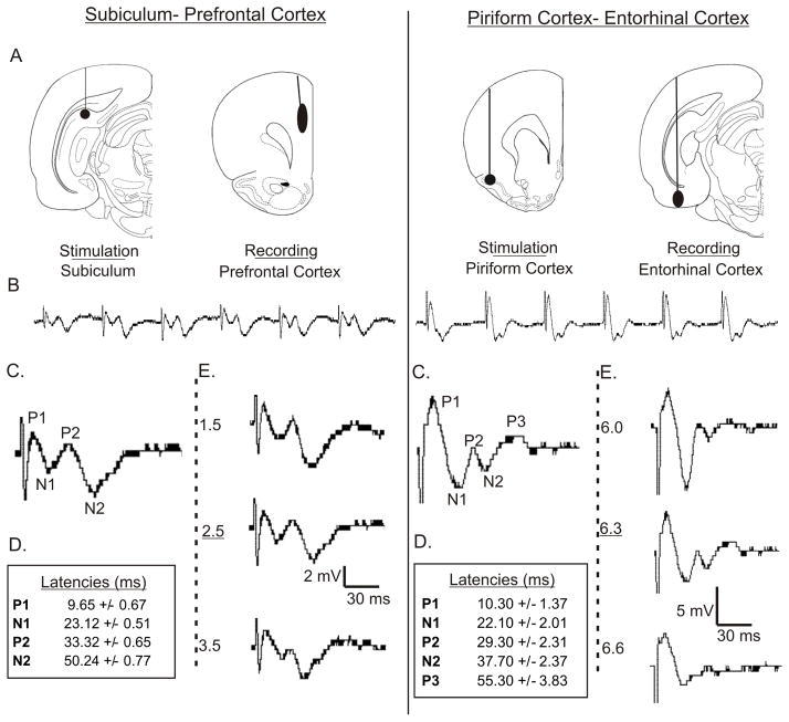
Introduction: The midline thalamic nuclei are an important component of limbic seizures. Although the anatomic connections and excitatory influences of the midline thalamus are well known, its physiological role in limbic seizures is unclear. We examined the role of the midline thalamus on two circuits that are involved in limbic seizures: (a) the subiculum-prefrontal cortex (SB-PFC), and (b) the piriform cortex-entorhinal cortex (PC-EC).

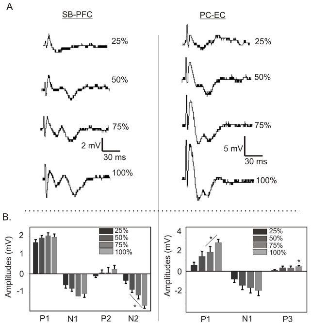
Methods: Evoked field potentials for both circuits were obtained in anesthetized rats, and the likely direct monosynaptic and polysynaptic contributions to the responses were identified. Seizures were generated in both circuits by 20 Hz stimulus trains. Once stable seizures and evoked potentials were established, the midline thalamus was inactivated through an injection of the sodium channel blocker tetrodotoxin (TTX), and the effects on the evoked responses and seizures were analyzed.

Results: Inactivation of the midline thalamus suppressed seizures in both circuits. Seizure suppression was associated with a significant reduction in the late thalamic component but no significant change in the early direct monosynaptic component. Injections that did not suppress the seizures did not alter the evoked potentials.

Conclusions: Suppression of the late thalamic component of the evoked potential at the time of seizure suppression suggests that the thalamus facilitates seizure induction by extending the duration of excitatory drive through a divergent-convergent excitatory amplification system. This work may have broader implications for understanding signaling in the limbic system.


Conflict of interest statement

The authors have no conflict of interest to declare.

Figures

Figure 1
Figure 1. Evoked Potentials From Two Limbic Circuits
(A) Electrode placement for SB-PFC and PC-EC responses (Brain section illustrations from Paxinos and Watson 1996). (B) Typical 120 ms i.s.i. train from the stimulation-recording pairs that shows the increasing amplitudes of the responses, plateauing by the 4th stimulation. (C) Typical 4th response from each train, with major peaks labeled. (D) Average latencies for each peak of the 4th responses. (E) Responses change with electrode depth. Position used in this study is underlined.
Figure 2
Figure 2. Effect of Increased Stimulation on Evoked Responses
To define maximal responses, as well as to determine the relative stimulus intensity needed to elicit each wave in the responses of both SB-PFC and PC-EC circuits, stimulus intensity was increased stepwise until maximum response. (A) Change of morphology and peak onset as intensities are increased, by increments of 25% of maximal stimulation. Note that responses at 50% intensity are different than those at 100%, and that all peaks are fully formed at maximum intensity. (B) Average peak amplitudes as intensities are increased. (* P< .05 by student’s t-test compared to 25% intensity).
Figure 3
Figure 3. Long-Term Potentiation in Both Circuits Identifies Early Monosynaptic Component
(A) 2nd responses from paired pulses in both pathways, taken at half-maximal intensity, before and after LTP was induced. Because of the low stimulation intensity, the peaks shown are not directly comparable to those in Figure 1. An overlay of the waveforms shows that the first primary negative wave has increased in amplitude. (B) Amplitudes of the positive and negative peaks. In both cases, the negative peak increases significantly (* P< .05 by student’s t-test). (C) Changes in the amplitude of the negative peak over the course of the experiment. Note the rise in amplitude after the LTP stimuli were given. The latency of these peaks occurs earlier than the probable thalamic component of the response, and fall within the range of the monosynaptic component (see Figure 4).
Figure 4
Figure 4. Estimation of Onset of Thalamic Component
To determine the approximate minimum time for the appearance for thalamic influence responses in the (A) SB-PFC and (B) PC-EC pathways, responses from the initiating site to the thalamus and the from the thalamus to the target site were obtained. Responses are aligned (1–3) and their latencies added to arrive at a minimum latency for thalamic involvement. The first major peak is treated as the monosynaptic response. The gradient bar (4) beneath the direct responses is placed to suggest that the onset of polysynaptic components occurs gradually within that suggested timeframe. The individual and combined latencies are quantified in the table inset. This figure is to be used and interpreted as a guide, and not an exact demonstration of thalamic influence in the direct response.
Figure 5
Figure 5. Limbic Seizures Affected by TTX in Midline Thalamus
(A) Limbic seizure afterdischarges before and after injection of TTX into the midline thalamus. Note that seizure activity is greatly reduced with TTX injection in both sets of responses. (B.) Average seizure durations before, during and after TTX with subsequent recovery. (C) Five-second samples from each trace (the boxed area on the full traces).
Figure 6
Figure 6. Evoked Potentials Affected by TTX in Midline Thalamus
(A) Sample averaged responses are shown for the SB-PFC (n=6) and PC-EC (n=5) circuits (3rd and 4th stimulations), before injection of TTX into the midline thalamus, after injection and after recovery. In both cases, the late component of the response is reduced in amplitude, and then recovers. (B) Amplitude differences between the peaks in the early (P1-N1) and late (P2-N2 in SB-PFC, P3-Baseline for PC-EC) components of the waveforms from the 4th stimulation in both pathways, and how they change with TTX injection in the thalamus. In both cases, the changes in the late components of the response are significantly reduced. *P<.05 by paired t-test.
Figure 7
Figure 7. Position Specificity of Effect of TTX on Seizures and Evoked Potentials
To show that the effects shown are specific to TTX injections in the mediodorsal nucleus, injections within and without that region are compared. (A) Both effective (filled circles) and ineffective (open circles) injections of TTX into the area of the midline thalamus are shown for both SB-PFC and PC-EC pathways. (Brain section illustrations from Paxinos and Watson 1996). (B) Sample seizure from an ineffective injection outside of the mediodorsal nucleus, showing a seizure before injection, and 30 minutes post injection. No change is duration is seen. (C) Evoked responses corresponding to the seizures shown, with no change seen. (D) Summary of average seizure durations and evoked potential peak amplitudes in both sets of animals with injections outside the MD (n=5 for SB-PFC, n=6 for PC-EC).
Figure 8
Figure 8. Representation of Divergent-Convergent Excitatory Circuits in the Limbic System
Model of a proposed role for the thalamus in limbic seizure circuits. A seizure is initiated in a region in the network (focus). Excitatory activity affects signaling to all the monosynaptic afferent targets of the region, including the midline thalamus. The thalamus, in turn, sends an additional, excitatory signal to all of the monosynaptic targets of the focus. This additional excitation assists in recruiting and driving the target structures into seizure activity by prolonged excitation.

References

    1. Avoli M, Gloor P. Interaction of cortex and thalamus in spike and wave discharges of feline generalized penicillin epilepsy. Exp Neurol. 1982;76:196–217. - PubMed
    1. Bazhenov M, Timofeev I, Steriade M, Sejnowski TJ. Cellular and network models of for intrathalamic augmenting responses during 10 Hz. stimulation. J Neurophysiol. 1998;79:2730–2748. - PubMed
    1. Berendse HW, Groenewegen HJ. Restricted cortical termination fields of the midline and intralaminar thalamic nuclei in the rat. Neuroscience. 1991;42:73–102. - PubMed
    1. Bertram EH. Functional anatomy of spontaneous seizures in a rat model of limbic epilepsy. Epilepsia. 1997;38:95–105. - PubMed
    1. Bertram EH, Zhang DX, Mangan P, Fountain N, Rempe D. Functional anatomy of limbic epilepsy: a proposal for central synchronization of a diffusely hyperexcitable network. Epilepsy Res. 1998;32:194–205. - PubMed

Publication types