Review
. 2011 May 12;70(3):385-409.
doi: 10.1016/j.neuron.2011.03.024.

GABAA receptor trafficking-mediated plasticity of inhibitory synapses

Affiliations
Review

GABAA receptor trafficking-mediated plasticity of inhibitory synapses

Bernhard Luscher et al. Neuron. .

Abstract

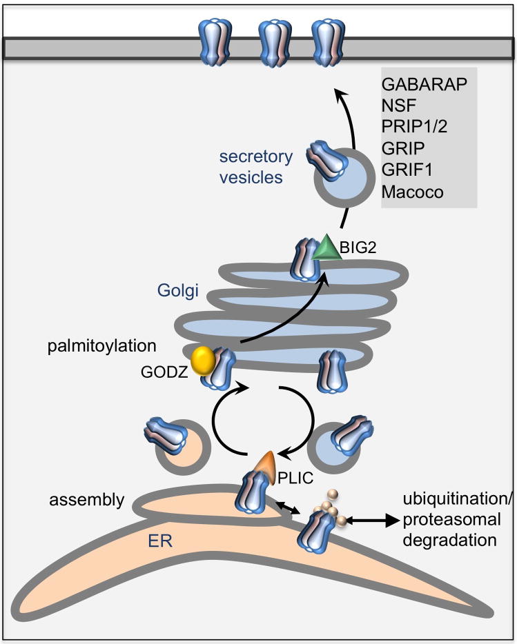
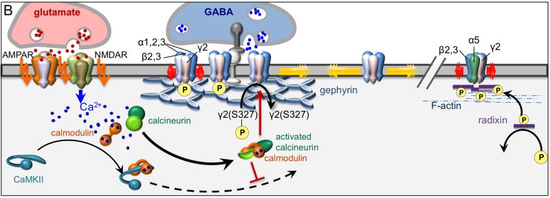
Proper developmental, neural cell-type-specific, and activity-dependent regulation of GABAergic transmission is essential for virtually all aspects of CNS function. The number of GABA(A) receptors in the postsynaptic membrane directly controls the efficacy of GABAergic synaptic transmission. Thus, regulated trafficking of GABA(A) receptors is essential for understanding brain function in both health and disease. Here we summarize recent progress in the understanding of mechanisms that allow dynamic adaptation of cell surface expression and postsynaptic accumulation and function of GABA(A) receptors. This includes activity-dependent and cell-type-specific changes in subunit gene expression, assembly of subunits into receptors, as well as exocytosis, endocytic recycling, diffusion dynamics, and degradation of GABA(A) receptors. In particular, we focus on the roles of receptor-interacting proteins, scaffold proteins, synaptic adhesion proteins, and enzymes that regulate the trafficking and function of receptors and associated proteins. In addition, we review neuropeptide signaling pathways that affect neural excitability through changes in GABA(A)R trafficking.


Conflict of interest statement

Conflicts of Interest. The authors declare no con ict of interest

Figures

Figure 1
Figure 1. GABAAR subunit structure and intracellular loop sequences
A. Schematic representation of GABAAR heteropentamers consisting of two α, two β and a single γ2 subunit assembled in a counterclockwise γ–β-α-β-α arrangement. B. Every subunit includes an extracellular N-terminal domain, four transmembrane domains (TM1–4) separated by an extended cytoplasmic loop region between TM3 and TM4, and a short extracellular C-terminus. The cytoplasmic loop and TM4 regions of the γ2 subunit are essential for postsynaptic clustering of GABAARs (Alldred et al., 2005). C. Sequences of cytoplasmic loop regions of representative subunits (γ2L, β3, α2) with amino acid numbers referring to mature polypeptides from the mouse. Interaction sites for binding partners are marked by brackets beneath the sequence, along with amino acid numbers of known Ser/Thr and Tyr phosphorylation sites. Phosphorylation sites are shown in blue, Lys residues representing putative ubiquitination sites are in orange. Note that the minimal interaction site for CAML includes the C-terminal region of the cytoplasmic loop as well as the TM4 domain of the γ2 subunit. For CaMKII phosphorylation sites see Houston et al (2009). For other references see text.
Figure 2
Figure 2. Regulated trafficking of GABAARs in the secretory pathway
GABAAR heteropentamers assemble in the ER and interact with chaperones including calnexin and BiP. Unassembled or improperly folded receptor subunits are subject to ubiquitination and proteasomal degradation. This process is inhibited by interaction of α and β subunits with the ubiquitin-like protein PLIC, which in turn promotes the exit of receptors from the ER to the Golgi. The Golgi resident palmitoyltransferase GODZ palmitoylates the receptor γ2 subunit at cytoplasmic cysteine residues, which promotes translocation of receptors through the Golgi apparatus to the plasma membrane and to synapses. Exit of GABAARs from the Golgi may be facilitated by interaction of the GTP exchange factor BIG2 with GABAAR β subunits. The surface delivery of GABAARs is further promoted by a number of other proteins that currently cannot be assigned to a specific trafficking compartment (shaded gray).
Figure 3
Figure 3. Schematic representation of proteins pivotal for intracellular trafficking of postsynaptic GABAARs
A. PRIP consists of an N-terminal domain that incudes a binding site for the catalytic domain of PP1α, a PH domain that includes a binding site for IP3 (D-myo-inositol 1,4,5 triphosphate), an EF-hand domain that includes a binding site for GABARAP, and homologies to the catalytic (X, Y) and C2 domains of phospholipase Cδ. The GABAAR β subunit interaction domain is located between the X and Y domains of PRIP. Amino acid numbers refer to murine PRIP-1 (Kanematsu et al., 2005). B. Gephyrin consists of an N-terminal G-domain involved in the formation of gephyrin trimers, a central C domain with interaction sites for Pin1, DLC1/2, collybistin, and a GSK3β phosphorylation site that regulates susceptibility to cleavage by calpain-1, and a C-terminal E domain that dimerizes in vitro and regulates clustering in vivo. In vitro assays suggest that the C domain also interacts with GABARAP and tubulin, although these proteins are not colocalized with postsynaptic gephyrin. The E domain binds Mena/VASP, profilin, the glycine receptor β subunit and neuroligins. The gephyrin motif at the C-E domain interface that interacts with collybistin (PFPLTSMDKA) (Harvey et al., 2004) overlaps with the α2 subunit binding site (SMDKAFITVLEMPTVLGTE) (Saiepour et al., 2010). Amino acid numbers refer to rat gephyrin (Prior et al., 1992). C. Collybistin exists in three alternatively spliced versions that differ in sequence and length of their C-terminal domain (striped area). In addition, its clustering function is regulated by the presence or absence of an SH3 domain. Also shown are the dbl homology (DH) domain that regulates nucleotide exchange and the pleckstrin homology (PH) domain required for interaction with membrane phosphoinositides. The gephyrin binding site has been mapped to the linker domain between SH3 and DH domains. Amino acid numbers refer to rat collybistin (Kins et al., 2000). D. NL2 is composed of an N-terminal signal peptide (SP), followed by a large choline-esterase-like domain with the alternatively spliced exon A, a transmembrane domain (TM), and an intracellular cytoplasmic domain that includes a 15-amino-acid tyrosine-containing binding site for gephyrin and a C-terminal binding site for PDZ domain proteins such as S-SCAM. Amino acid numbers refer to mouse NL2 (Ichtchenko et al., 1996).
Figure 4
Figure 4. Regulated endocytosis and recycling of GABAARs
GABAAR endocytosis through clathrin-coated vesicles is regulated by phospho-sensitive interactions of β and γ2 subunits with the clathrin adaptor AP2. Phosphorylation of β subunits (S408/409 in β3) and the γ2 subunit (Y365/367) by PKA/PKC and Fyn/Src, respectively interferes with these interactions and thereby stabilizes GABAARs at the cell surface. Phosphorylation of β subunits by PKA and PKC is facilitated by the kinase adaptors AKAP and RACK, respectively. Dephosphorylation is modulated by PRIP-associated PP1α and PP2A. GABAARs in the plasma membrane are subject to lateral diffusion. Interaction of GABAARs with gephyrin (i.e. through α2/3 subunits) and collybistin (α2 subunit) leads to their accumulation at synapses. Interaction of gephyrin/collybistin/GABAAR complexes with the NL2-neurexin synaptic adhesion complex contributes to proper alignment of pre- and postsynaptic complexes at inhibitory synapses. Conversely, dephosphorylation of β subunits by PP1α and PP2A (β3 subunit S408/409 site) and unidentified tyrosine phosphatases (γ2 Y365/367) facilitates interaction of extrasynaptic GABAARs with AP2, which then triggers clathrin-mediated internalization. Dephosphorylation of β subunits by PP1α is inhibited or facilitated by the phosphatase adaptor PRIP, depending on its own phosphorylation state. Endocytosed receptors in early endosomes are ubiquitinated at lysine and possibly other residues of the γ2 subunit, which then leads to lysosomal degradation. Alternatively, interactions of CAML with the γ2 subunit cytoplasmic and transmembrane domains and of HAP1 with the β subunit cytoplasmic domain facilitate KIF5-dependent vesicular transport and recycling of GABAARs to the plasma membrane.
Figure 5
Figure 5. Regulation of GABAAR clustering and lateral mobility at synaptic and extrasynaptic sites
A. The biosynthesis of gephyrin is regulated by the peptidyl-prolyl cis/trans isomerase Pin1. Cytosolic soluble gephyrin exists as a trimer. The deposition of gephyrin trimers at the plasma membrane is facilitated by cooperative interactions of gephyrin with CBSH3+ (tethered to the plasma membrane by phosphoinositide binding of its PH domain) and NL2, which unlock the CBSH3+-dependent clustering function, presumably by releasing an intramolecular inhibition of CBSH3+ by its SH3 domain. The gephyrin/NL2/collybistin complex enables the postsynaptic clustering of gephyrin and, through interaction with presynaptic neurexins, helps to align the postsynaptic complex with GABAergic terminals. The GABAAR α2 subunit may substitute for NL2 and enable collybistin-dependent clustering of gephyrin. The clustering of GABAARs in the postsynaptic specialization is facilitated by interaction of specific subunits (α2, α3) with gephyrin. Postsynaptic gephyrin further interacts with Mena/VASP and profilin I/II. Competition of gephyrin and G-actin for interaction with profilin I/II is implicated in regulation of the microfilament-dependent receptor packing density. The density of postsynaptic gephyrin clusters is regulated by GSK3β-mediated phosphorylation of gephyrin, which enhances the susceptibility of gephyrin to cleavage by the Ca2+-dependent protease calpain-1. Constitutive proteolytic cleavage of gephyrin limits the confinement and accumulation of postsynaptic GABAARs, by facilitating their lateral diffusion. Conversely, inhibition of GSK3β by Li+ or of calpain-1 by its natural antagonist calpastatin stabilizes gephyrin and thereby promotes the density of gephyrin clusters as well as GABAergic synaptic function. B. Postsynaptic GABAARs (α1,2,3β2/3γ2) are confined (red arrows) by interactions with gephyrin and presumably other postsynaptic scaffold proteins. However, on leaving this area they become highly mobile within the plane of the phospholipid bilayer (yellow arrows). The interaction of GABAARs with the postsynaptic cytoskeleton is regulated by the activity-dependent and calcineurin-regulated phosphorylation state of the γ2 subunit. Robust excitation of glutamate receptors leads to NMDAR/Ca2+- and Ca2+/calmodulin-mediated activation of calcineurin and dephosphorylation of γ2(S270), which reduces the postsynaptic confinement of GABAARs allowing their diffusion away from synapses. CaMKII is activated in parallel but its translocation to synapses is prevented by calcineurin by an unknown mechanism. In contrast to α1,2,3β2/3γ2 receptors, α5βγ2 receptors are clustered extrasynaptically by interaction with phospho-activated radixin, which links these receptors to submembrane microfilaments. C. Modest stimulation of neurons as mimicked by treatment of neurons with NMDA leads to more limited influx of Ca2+ and preferential activation of CaMKII. Activated CaMKII is translocated to synapses and stimulates the insertion of GABAARs into the plasma membrane where they are trapped at synapses by interaction with the postsynaptic cytoskeleton. Insertion of GABAARs into the plasma membrane involves GABARAP, NSF and GRIP. The relevant target proteins interacting with and phosphorylated by CaMKII are not yet known.
Figure 6
Figure 6. Insulin receptor-mediated surface expression of GABAARs
Two distinct mechanisms have been proposed for insulin-induced surface expression of GABAARs that involve either Ser or Tyr phosphorylation of the GABAAR β subunit, respectively and may function independently or cooperatively. A. In a first mechanism, insulin receptor-mediated phosphorylation of the insulin receptor substrate-1 (IRS-1) leads to activation of PI3K and accumulation of phosphoinositide (PIP3) at the plasma membrane. PIP3-mediated recruitment of Akt to the plasma membrane facilitates PDK1-mediated phosphorylation and activation of Akt. Phosphorylated Akt forms a ternary complex with PRIP and vesicular GABAARs, which causes phosphorylation (β3S408/9) and translocation of GABAARs to the plasma membrane. As an additional primary downstream target of insulin, Akt is known to inhibit GSK3β. This kinase in turn phosphorylates gephyrin, which triggers calpain-1-mediated degradation of gephyrin. Thus, insulin signaling may promote GABAergic synaptic function by increasing the surface expression and gephyrin-dependent synaptic confinement of GABAARs. B. In an alternate mechanism, the PI3K P85 subunit interacts directly with GABAARs. This complex is abundant already under basal condition and dependent on phosphorylated Tyr residues (Y372/9) of the GABAAR β3 subunit. On stimulation with insulin, the abundance of this complex and its association with phosphorylated lipids (PIP3) is rapidly increased, concurrent with translocation of the complex to the plasma membrane.

Similar articles

Cited by

References

    1. Abrami L, Kunz B, Iacovache I, van der Goot FG. Palmitoylation and ubiquitination regulate exit of the Wnt signaling protein LRP6 from the endoplasmic reticulum. Proc Natl Acad Sci U S A. 2008;105:5384–5389. - PMC - PubMed
    1. Abramian AM, Comenencia-Ortiz E, Vithlani M, Tretter EV, Sieghart W, Davies PA, Moss SJ. Protein kinase C phosphorylation regulates membrane insertion of GABA(A) receptor subtypes that mediate tonic inhibition. J Biol Chem. 2010;285:41795–41805. - PMC - PubMed
    1. Alldred MJ, Mulder-Rosi J, Lingenfelter SE, Chen G, Luscher B. Distinct gamma2 subunit domains mediate clustering and synaptic function of postsynaptic GABAA receptors and gephyrin. J Neurosci. 2005;25:594–603. - PMC - PubMed
    1. Alle H, Geiger JR. GABAergic spill-over transmission onto hippocampal mossy fiber boutons. J Neurosci. 2007;27:942–950. - PMC - PubMed
    1. Andang M, Lendahl U. Ion fluxes and neurotransmitters signaling in neural development. Current opinion in neurobiology. 2008;18:232–236. - PubMed

Publication types

Substances

LinkOut - more resources