. 2011 May 12;70(3):427-40.
doi: 10.1016/j.neuron.2011.03.021.

An antisense CAG repeat transcript at JPH3 locus mediates expanded polyglutamine protein toxicity in Huntington's disease-like 2 mice

Affiliations

An antisense CAG repeat transcript at JPH3 locus mediates expanded polyglutamine protein toxicity in Huntington's disease-like 2 mice

Brian Wilburn et al. Neuron. .

Abstract

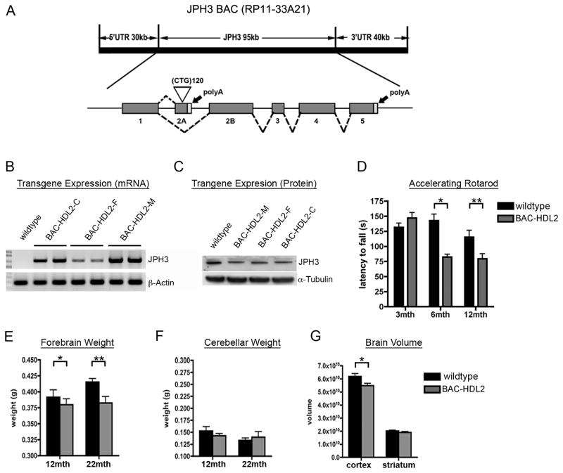
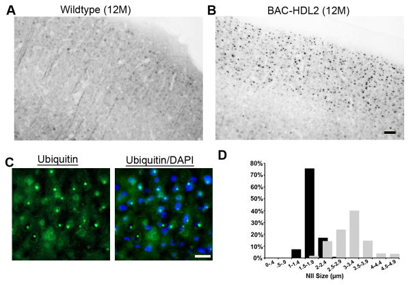
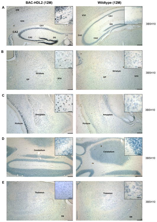
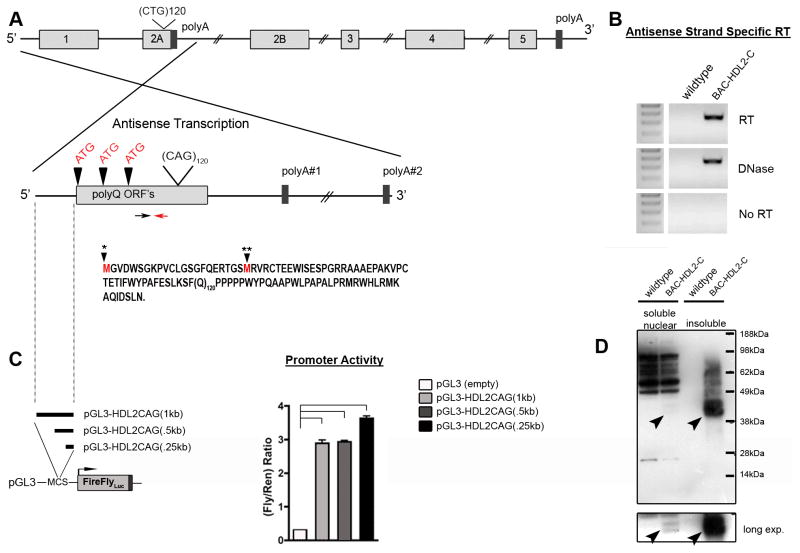
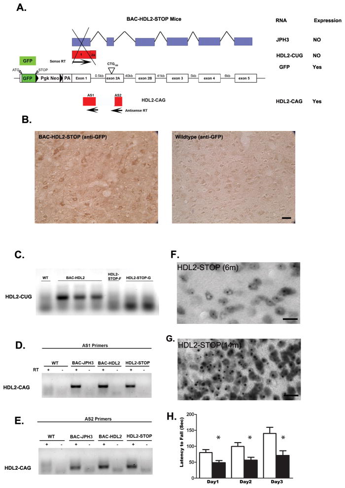
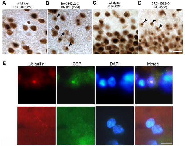
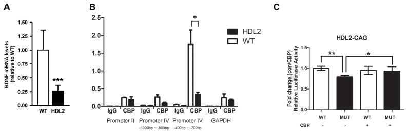
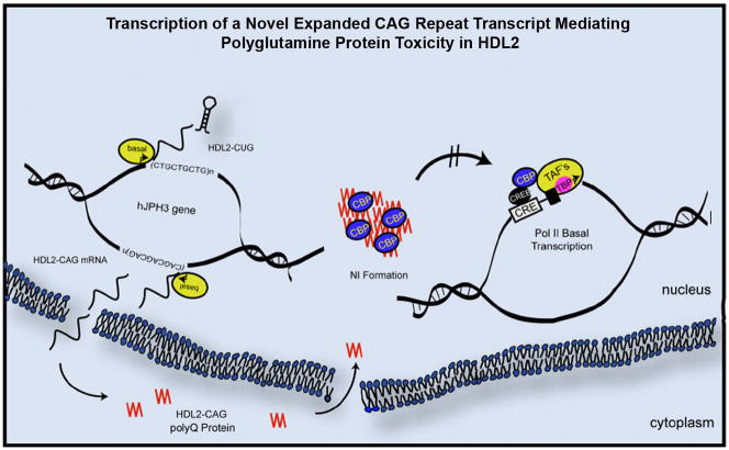
Huntington's disease-like-2 (HDL2) is a phenocopy of Huntington's disease caused by CTG/CAG repeat expansion at the Junctophilin-3 (JPH3) locus. The mechanisms underlying HDL2 pathogenesis remain unclear. Here we developed a BAC transgenic mouse model of HDL2 (BAC-HDL2) that exhibits progressive motor deficits, selective neurodegenerative pathology, and ubiquitin-positive nuclear inclusions (NIs). Molecular analyses reveal a promoter at the transgene locus driving the expression of a CAG repeat transcript (HDL2-CAG) from the strand antisense to JPH3, which encodes an expanded polyglutamine (polyQ) protein. Importantly, BAC-HDL2 mice, but not control BAC mice, accumulate polyQ-containing NIs in a pattern strikingly similar to those in the patients. Furthermore, BAC mice with genetic silencing of the expanded CUG transcript still express HDL2-CAG transcript and manifest polyQ pathogenesis. Finally, studies of HDL2 mice and patients revealed CBP sequestration into NIs and evidence for interference of CBP-mediated transcriptional activation. These results suggest overlapping polyQ-mediated pathogenic mechanisms in HD and HDL2.


Figures

Figure 1
Figure 1
Generation and Characterization of BAC-HDL2 animals. (A) A schematic representation of the human JPH3 locus. A BAC containing the intact JPH locus (RP11-33A21) was modified in order to insert ~120 CTG repeats into the alternatively spliced exon 2A (white triangle). (B) RT-PCR performed using primers specific to human JPH3 exons 2B and 4. (C) JPH3 Western blot performed on brain lysates isolated from BAC-HDL2C/M/F lines and wildtype control. (D) Accelerating rotarod testing performed at 3, 6 and 12 months of age (3 month: wild-type N=22, BAC-HDL2-C N=16; 6 month: wild-type N=14, BAC-HDL2-C N=11; 12 month: wild-type N=10, BAC-HDL2-C N=11). (E) BAC-HDL2-C line forebrains weigh significantly less than their wild-type littermates at both 12 and 22 months of age. (F) No significant differences detected in cerebellum weights between HDL2-C and its wild type littermates at 12 and 22 months. (G) Stereological brain volume measurements of BAC-HDL2-C line cortex and striatum revealed a significant decrease in the cortical volume at 18–22 months of age. (BAC-HDL2-C: striatum, N=6, cortex, N=6; wildtype, striatum, N=6, cortex, N=6). Values are mean ± SEM (*p< 0.05, **p< 0.01, Student’s t test).
Figure 2
Figure 2
Ubiquitin immunoreactive nuclear inclusions in BAC-HDL2 brains. (A,B) Antigen retrieval followed by immunohistochemical staining with 1C2 antibody. 12 month old BAC-HDL2-C brain sections revealed numerous large inclusion bodies in the cortical layers II/III (B). No such staining was observed in the respective brain regions in the wildtype animals (A); (C) Ubiquitin immunoreactive inclusion bodies in BAC-HDL2-C brain are co-localized with the nuclear marker DAPI, hence they are nuclear inclusions (NIs). (D) Quantification of NI size at 3 and 12 months of age with summary histograms shown [mean=1.8 μm vs. 3.13 μm, for 3 and 12 months respectively; *p<.001, Student’s t test; BAC-HDL2-C: motor sensory (MS) cortex layers I/II/III, 3 month, (155 NIs measured in two mice), 12 month (194 NIs measured in two mice)]. Scale bar, 50 μm.
Figure 3
Figure 3
The distribution of 3B5H10 immunoreactive NIs in BAC-HDL2 brains recapitulates that seen in HDL2 patients. Immunostaining with a monoclonal antibody against expanded polyQ (3B5H10) in 12 month old BAC-HDL2 and wildtype brain sections. Prominent 3B5H10 stained aggregates are detected in the mutant but not wildtype mouse brains in the upper cortical layers and hippocampus (A), striatum (B), amygdala (C). Very few if any 3B5H10 immunoreactive aggregates were detected in the cerebellum (D) or thalamus (E). Scale bar, 200 μm; Inset scale bar, 25 μm.
Figure 4
Figure 4
Expression of mutant HDL2-CAG RNA and proteins in BAC-HDL2 brains. (A) Graphic representation of potential HDL2-CAG transcript(s) containing the expanded CAG repeat with polyQ ORF’s (vertical arrowheads). The two closest polyA signals are shown. A red arrow marks the location of the primer used to drive reverse-strand-specific cDNA synthesis. The black arrow marks the location of the forward primer used for PCR after cDNA synthesis. Defined HDL2-CAG mutant transcripts include two potential ORF’s through the CAG repeat (black arrowhead with star marks) based on the differential usage of the two ATG translational initiation codons that can be amplified (the first ATG codon is not in any 5′ RACE product); (B) RT-PCR analyses provide evidence for the expression of HDL2-CAG transcripts emanating from the JPH3 antisense strand in BAC-HDL2-C animals. (C) DNA upstream of HDL2-CAG (position defined by dotted line) was cloned in front of luciferase in order to detect promoter activity in primary cortical neurons (*p<0.01 for pGL3-HDL2CAG1kb, -HDL2CAG.5kb and -HDL2CAG.25kb compared to empty vector (pGL3-empty); values are mean ± SEM). (D) Western blot analysis using 3B5H10 antibody was performed on soluble cytoplasmic and nuclear soluble fractions (i.e. cyto sol or nuc sol) as well as insoluble cellular fractions (i.e. insol; crude inclusion fraction) extracted from the forebrain of BAC-HDL2-C and wildtype mice at 18 months of age. Black arrowheads point to novel disease associated polyQ protein bands found specifically in BAC-HDL2-C line brains but not in wildtype littermate controls. This novel polyQ protein (termed HDL2-CAG) markedly accumulates in the insoluble fraction in the aged mutant brains.
Figure 5
Figure 5
Antisense HDL2-CAG transcript and accumulation of polyQ are independent of the expression of sense strand of JPH3 gene. (A) A graphic representation of the construct of BAC-HDL2-STOP mice. A transcriptional STOP sequence consisted of GFP followed by triple polyadenylation signal was placed in front of the translation initiation codon of JPH3 in the exon1 of the JPH3 on the BAC transgene. This mouse line was designed to express GFP from JPH3 promoter, but there is no transcription and translation of any other mRNA in the JPH3 sense strand, including any of the transcripts that include the expanded CUG repeat. (B) Anti-GFP antibody readily detected the expression of GFP protein in the cortical neurons in BAC-HDL2-STOP mice but not WT controls. (C). JPH3-sense-strand specific RT-PCR to amplify HDL2-CUG transcript revealed the expression of such transcript in BAC-HDL2-C mice, but not in two lines of BAC-HDL2-STOP mice (lines F and G). (D) & (E). Two independent strand-specific RT-PCR primer sets (AS1 and AS2 primers) specific to the HDL2-CAG transcripts readily detected the antisense CAG transcript in JPH3-FvB control mice, BAC-HDL2 mice, and BAC-HDL2-STOP mice. (F and G). 3B5H10 immunostaining reveals progressive accumulation of polyQ NIs in cortical neurons of BAC-HDL2-STROP mice at 6 and 12 months of age, but such NIs were not detected in the same brain region in the wildtype littermates (data not shown). (H). Rotarod deficits in 12 month old BAC-HDL2-STOP mice compared to wildtype controls. Significant deficits were detected at all three testing days using a Students test (with *, P< 0.05). Repeat-measure ANOVA analysis revealed a significant effect of time (F(2,8) = 9.250; p < 0.0001), genotype (F(2,8) = 9.331; p = 0.009), and interaction of time and genotype (F(2,8) = 3.026; p < 0.0001). Scale bar, 50 μm.
Figure 6
Figure 6
CBP sequestration in NIs in BAC-HDL2 Mice and HDL2 Patient Brains. (A–D) Two anti-CBP antibodies (A-22 and sc-583) were used to confirm the presence of CBP immunoreactive NIs in BAC-HDL2 mice (sc-583 is shown). Representative images of CBP staining of the superficial cortical layers (Cortical layers II/III; 6A and 6B) and hippocampal dentate gyrus (DG; 6C and 6D) for wildtype (6A and 6C) and BAC-HDL2 (6B and 6D) sections were shown. Selected NIs are highlighted with black arrowheads. (E) Double immunofluorescence staining using anti-CBP and anti-ubiquitin antibodies revealed the presence of CBP immunoreactive NIs that co-localize with ubiquitin staining in the nucleus (DAPI staining) in the cortical cells in HDL2 postmortem brain. Cortical neurons in control brains do not have CBP and ubiquitin-immunoreactive NIs, and CBP is diffusely distributed. Scale bar, 50 μm
Figure 7
Figure 7
CBP mediated transcriptional dysregulation in mouse and primary neuronal models of HDL2. (A) Quantitative RT-PCR analyses of the intact BDNF coding region reveal a significant reduction of BDNF transcripts in 15-month-old BAC-HDL2 mice relative to wildtype control mice (N=3 per genotype; ***p < 0.01, Student’s t-test). Values are mean ± SEM. (B). CBP occupancy of mouse BDNF promoter II, promoter IV (proximal region: 200–400bp from the transcription start site; distal region: 800–1000bp from the start site), BDNF coding region and GAPHD promoter were determined by ChIP-PCR in cortical samples derived from wildtype and BAC-HDL2 mice at 15 months of age (N=3 per genotype). ChIP-qPCR (IP/WCE,%) signals were normalized to whole cell extract (WCE). Immunoprecipitation with IgG was used for controls. Error bar represent S.E.M. determined from three independent experiments. The results suggest a significant and selective reduction of CBP binding to the proximal BDNF promoter IV (200–400bp) in cortical samples from mutant BAC-HDL2 mice compared to WT controls. Values are mean ± SEM (*p<0.01, Student’s t test). (C). Luciferase reporter assays were performed in rat cortical primary neurons with co-transfection of BDNF promoter IV-driven firefly luciferase reporter construct and vectors expressing either HDL2-CAG14 (wildtype HDL2-CAG protein with 14 glutamine repeat; labeled WT) or HDL2-CAG120 (mutant protein with 120 glutamine repeats; labeled HDL2). A separate set of transfection assays was performed with the same plasmids as above but the addition of plasmids overexpressing CBP (labeled CBP +). A renilla luciferase expression plasmid was used to normalize transfection efficiency in all the assays. Values are mean ± SEM (**p<0.01, *p<0.05, Student’s t test).
Figure 8
Figure 8
A schematic model for novel pathogenic mechanisms revealed by HDL2 mouse model in which a novel antisense expanded CAG transcript is mediating polyQ pathogenesis in vivo. A novel expanded CAG repeat (HDL2-CAG) antisense to the JPH3 sense strand is transcribed from its own promoter immediately 5′ to the CAG repeat, and the transcript is then translated into a novel expanded polyQ protein termed HDL2-CAG. Mutant HDL2-CAG is translocated and accumulated in the nucleus to form prominent NIs consisting of HDL2-CAG protein, ubiquitin and at a later time point, CBP. Transcriptional dysregulation, in part due to interference of CBP-mediated activation of gene expression (e.g. BDNF), may be a shared molecular pathogenic mechanism between HD and HDL2.

Comment in

References

    1. Brooks E, Arrasate M, Cheung K, Finkbeiner SM. Using antibodies to analyze polyglutamine stretches. Methods Mol Biol. 2004;277:103–128. - PubMed
    1. Chan HM, La Thangue NB. p300/CBP proteins: HATs for transcriptional bridges and scaffolds. J Cell Sci. 2001;114:2363–2373. - PubMed
    1. Dorsman JC, Pepers B, Langenberg D, Kerkdijk H, Ijszenga M, den Dunnen JT, Roos RA, van Ommen GJ. Strong aggregation and increased toxicity of polyleucine over polyglutamine stretches in mammalian cells. Hum Mol Genet. 2002;11:1487–1496. - PubMed
    1. Gambazzi L, Gokce O, Seredenina T, Katsyuba E, Runne H, Markram H, Giugliano M, Luthi-Carter R. Diminished activity-dependent brain-derived neurotrophic factor expression underlies cortical neuron microcircuit hypoconnectivity resulting from exposure to mutant huntingtin fragments. J Pharmacol Exp Ther. 2010;335:13–22. - PubMed
    1. Gong S, Yang XW, Li C, Heintz N. Highly efficient modification of bacterial artificial chromosomes (BACs) using novel shuttle vectors containing the R6Kgamma origin of replication. Genome Res. 2002;12:1992–1998. - PMC - PubMed

Publication types

MeSH terms