. 2011 May 12;70(3):468-81.
doi: 10.1016/j.neuron.2011.03.020.

Neurexin-neuroligin transsynaptic interaction mediates learning-related synaptic remodeling and long-term facilitation in aplysia

Affiliations

Neurexin-neuroligin transsynaptic interaction mediates learning-related synaptic remodeling and long-term facilitation in aplysia

Yun-Beom Choi et al. Neuron. .

Abstract

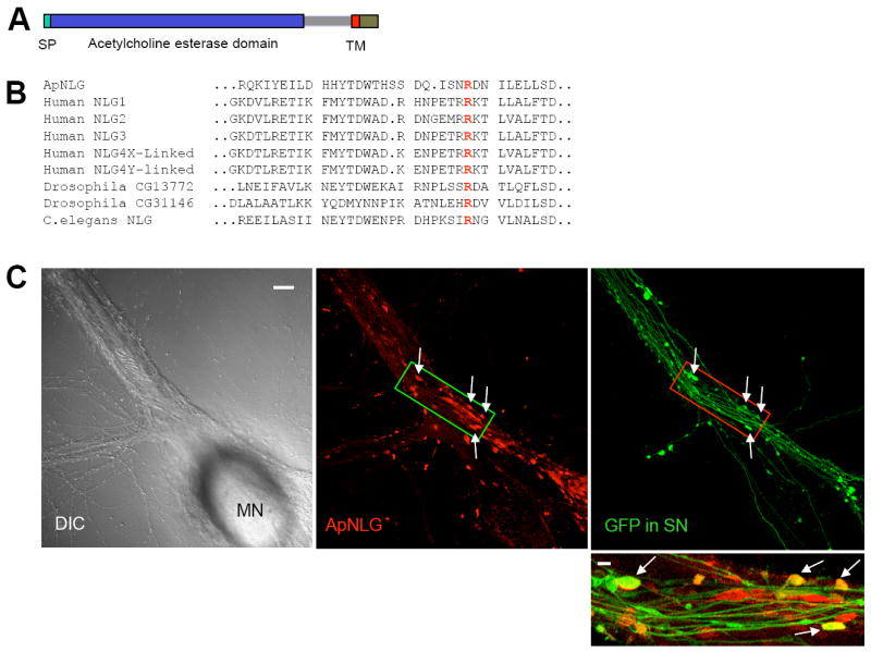
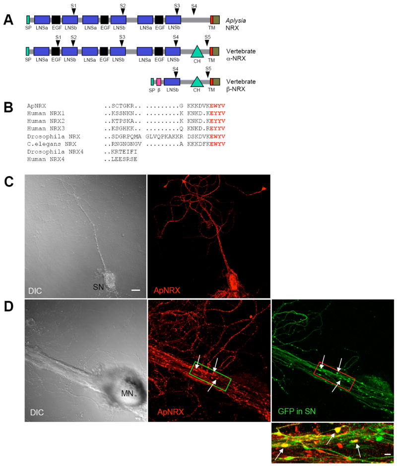
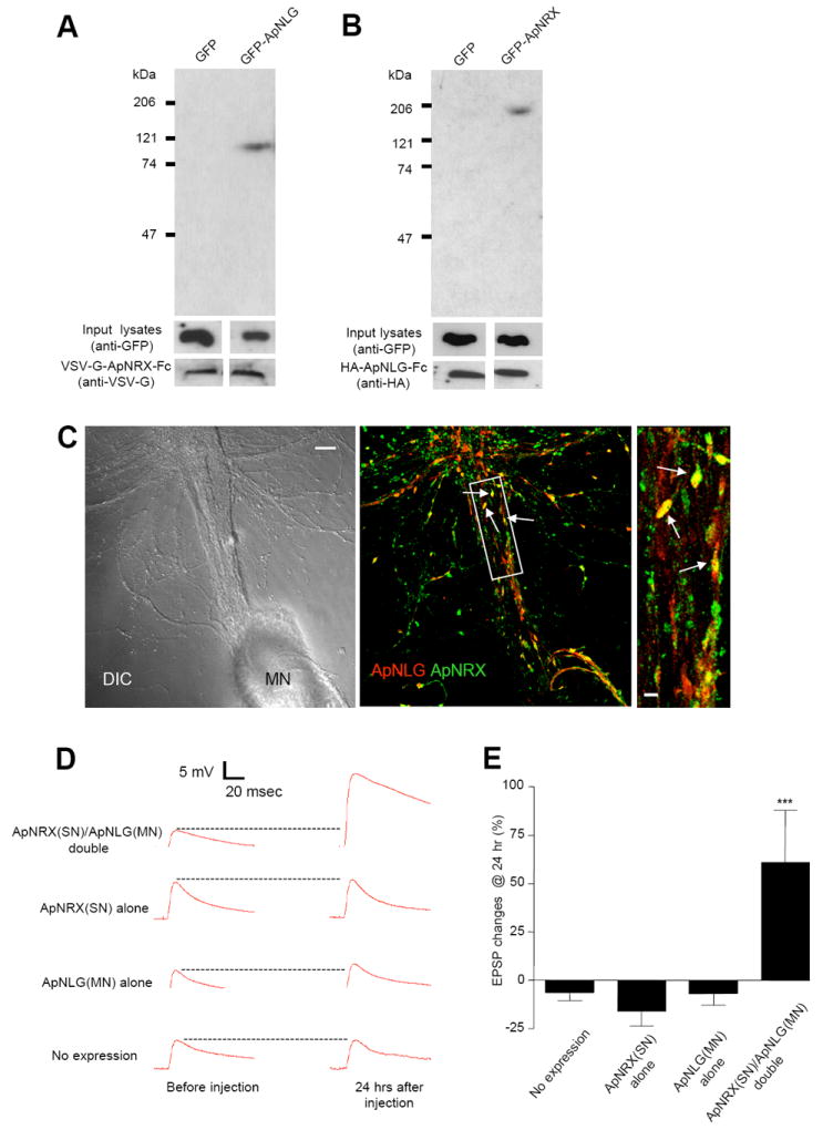
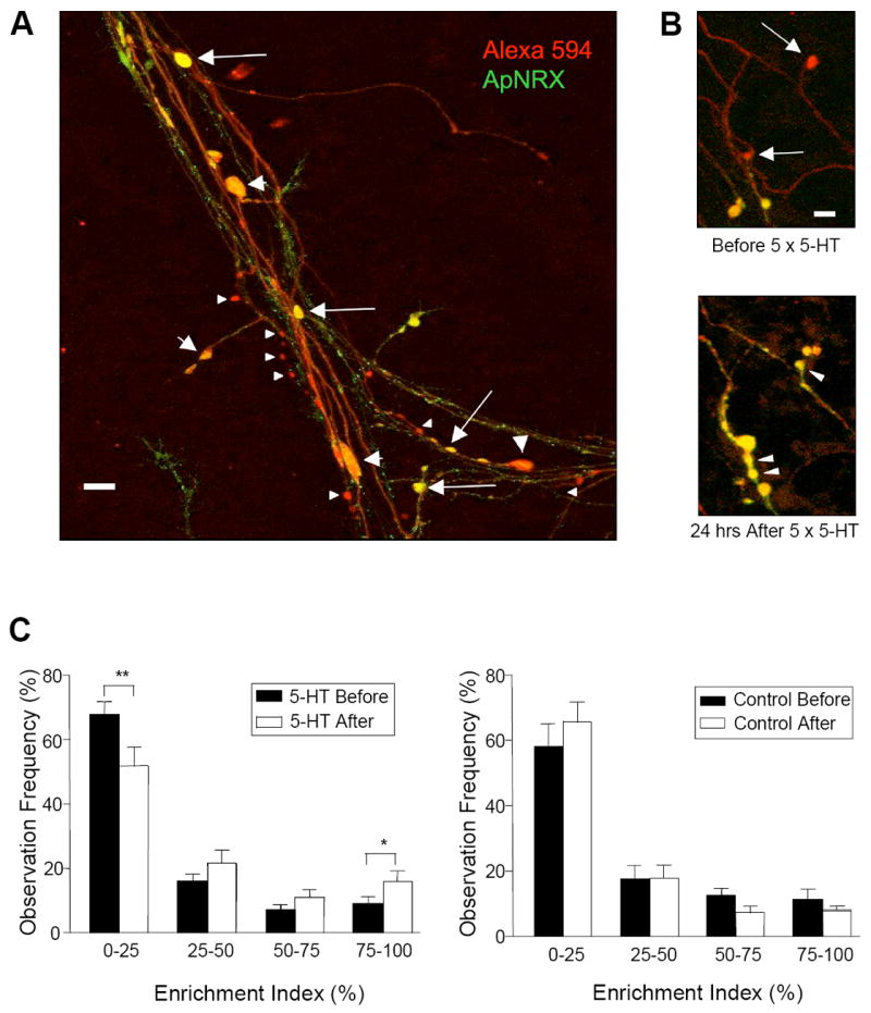
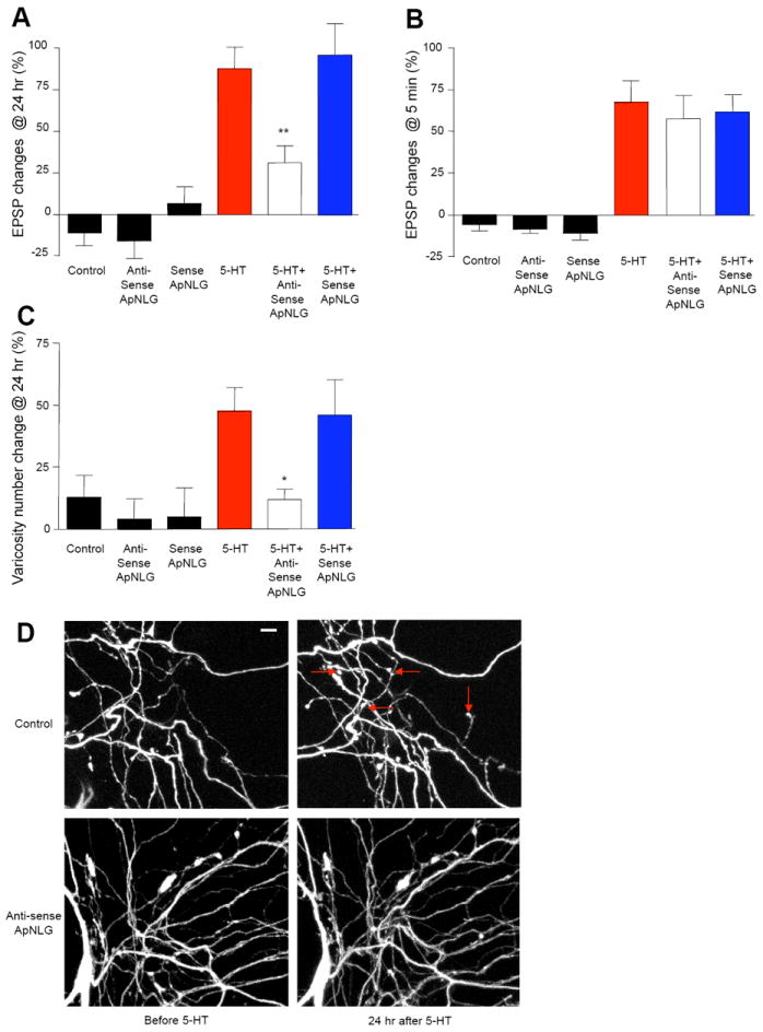
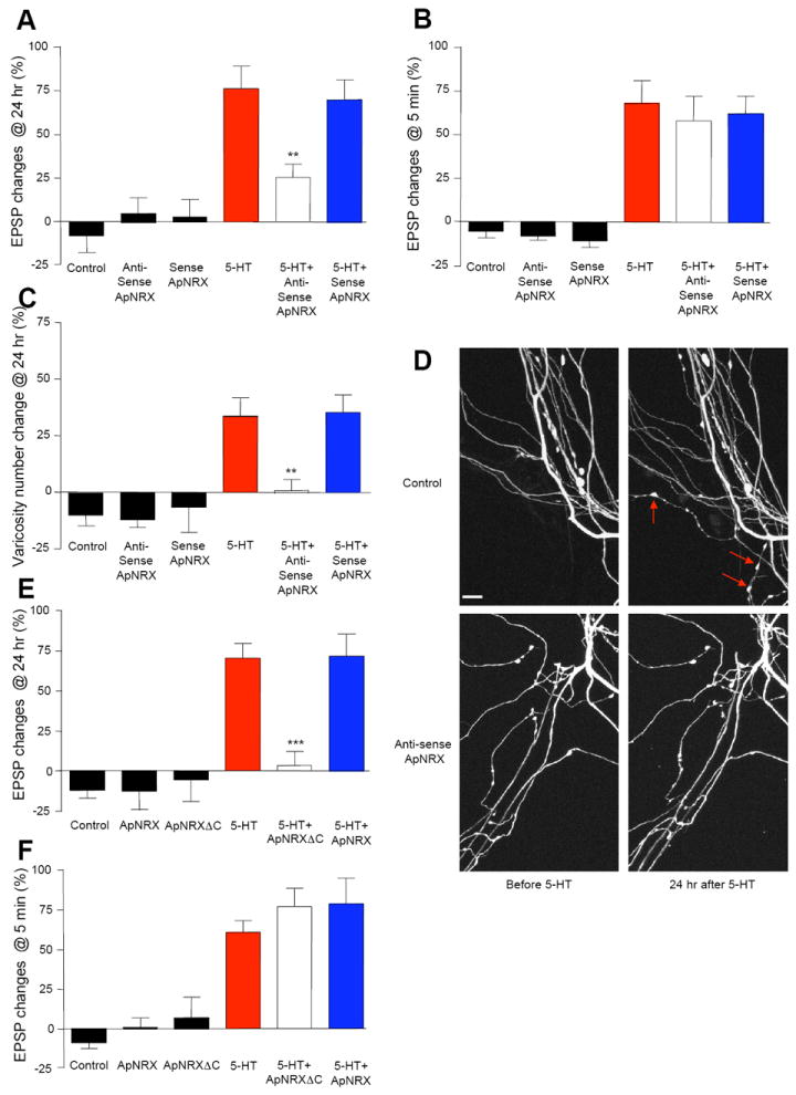
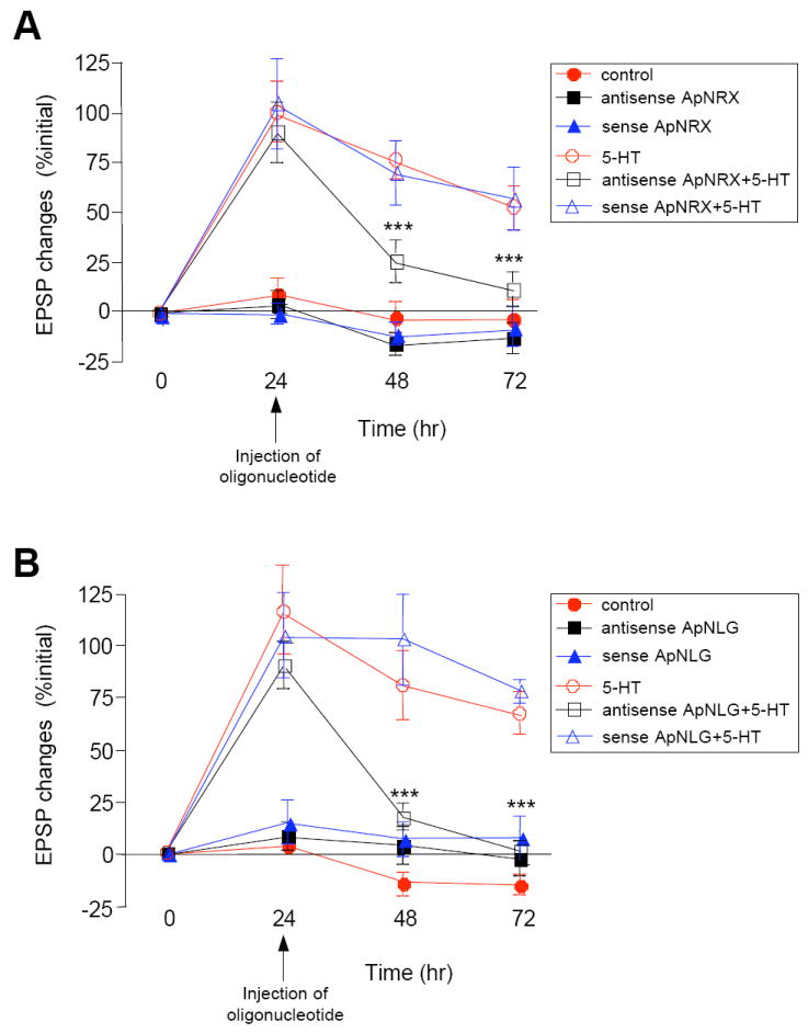
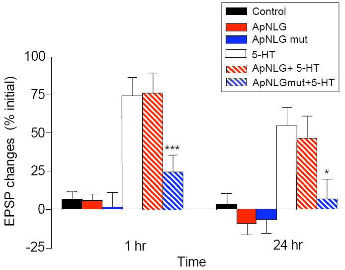
Neurexin and neuroligin, which undergo heterophilic interactions with each other at the synapse, are mutated in some patients with autism spectrum disorder, a set of disorders characterized by deficits in social and emotional learning. We have explored the role of neurexin and neuroligin at sensory-to-motor neuron synapses of the gill-withdrawal reflex in Aplysia, which undergoes sensitization, a simple form of learned fear. We find that depleting neurexin in the presynaptic sensory neuron or neuroligin in the postsynaptic motor neuron abolishes both long-term facilitation and the associated presynaptic growth induced by repeated pulses of serotonin. Moreover, introduction into the motor neuron of the R451C mutation of neuroligin-3 linked to autism spectrum disorder blocks both intermediate-term and long-term facilitation. Our results suggest that activity-dependent regulation of the neurexin-neuroligin interaction may govern transsynaptic signaling required for the storage of long-term memory, including emotional memory that may be impaired in autism spectrum disorder.


Figures

Figure 1
Figure 1. Cloning and Subcellular Localization of ApNLG
(A) The domain structure of ApNLG is similar to vertebrate neuroligin. SP= signal peptide. TM=transmembrane domain. (B) Comparison of the deduced amino acid sequence of ApNLG (partial) with vertebrate and invertebrate homologs. A highly conserved arginine (R) residue that is mutated in human NLG-3 R451C linked to ASD is present in the ApNLG (bold red). (C) Left: A DIC image shows a sensory-to-motor neuron co-culture. MN denotes the motor neuron cell body. Sensory neuron cell body is located outside of the field of view. Middle: ApNLG immunostaining (red) shows clustering of ApNLG at the initial segment and proximal regions of the major axons of the postsynaptic motor neuron. Right: GFP (green) as a whole cell marker outlines presynaptic sensory neuron processes and varicosities. Scale bar 20 μm. Bottom: A merged image in a magnified view shows some sensory neuron varicosities that partially or completely overlap with ApNLG clusters (yellow, arrows). Magnified view, scale bar 5 μm. See also Figure S1 and S2.
Figure 2
Figure 2. Cloning and Subcellular Localization of ApNRX
(A) The domain structure of ApNRX is similar to vertebrate α -neurexins. SP=signal peptide, β = β-neurexin-specific domain, LNSa and LNSb= LNS domain preceding and following EGF domain, respectively. CH=carbohydrate rich domain. TM=transmembrane domain. Arrowheads indicate splicing sites. (B) Comparison of the deduced amino acid sequence of the C-terminal end of ApNRX with vertebrate and invertebrate homologs. Bold red letters indicate the conserved residues in the C-terminal end PDZ domain. (C) Left: A DIC image of an isolated sensory neuron (SN). Right: ApNRX immunostaining (red) shows clustering of endogenous ApNRX along the neurites. (D) Left: A DIC image shows a sensory-to-motor neuron co-culture. MN denotes the motor neuron cell body. Sensory neuron cell body is located outside of the field of view. Middle: ApNRX immunostaining (red) shows clustering of ApNRX at the main neurites of postsynaptic motor neuron. Right: GFP (green) as a whole cell marker outlines presynaptic sensory neuron processes and varicosities. Scale bar 20 μm. Bottom: A merged image in a magnified view shows some sensory neuron varicosities that partially or completely overlap with ApNLG clusters (yellow, arrows). Magnified view, scale bar 5 μm. See also Figure S1 and S2 and Table S1.
Figure 3
Figure 3. Binding of Recombinant ApNRX with ApNLG and Enhancement of Basal EPSP by Simultaneous Overexpression of ApNRX in Presynaptic Sensory Neuron and ApNLG in Postsynaptic Motor Neuron
(A) Pull-down of GFP-ApNLG by VSV-G-ApNRX-Fc fusion protein. Immunoblotting with an anti-GFP antibody shows the presence of a 110 kDa band in the precipitate from HEK 293 cell lysates transfected with GFP-ApNLG. Similar amounts of input lysates and VSV-G-ApNLG-Fc fusion protein are shown when probed with an anti-GFP antibody and an anti-VSV-G antibody, respectively. (B) Pull-down of GFP-ApNRX by HA-ApNLG-Fc fusion protein. Immunoblotting with an anti-GFP antibody shows the presence of a 200 kDa band in the precipitate from HEK 293 cell lysates transfected with GFP-ApNRX. Similar amounts of input lysates and HA-ApNLG-Fc fusion protein are shown when probed with an anti-GFP antibody and an anti-HA antibody, respectively. (C) Left: A DIC image shows a sensory-to-motor neuron co-culture. MN denotes the motor neuron cell body. Sensory neuron cell body is located outside of the field of view. Middle: A merged co-immunostaining image shows clusters of ApNLG (red) and ApNRX (green) overlap or juxtapose at the initial segment and proximal regions of the major axons of the postsynaptic motor neuron (yellow, arrows). Scale bar 20 μm. Right: A magnified view, scale bar 5 μm. (D) EPSPs were recorded from motor neurons in response to extracellular stimulation of sensory neurons before and 24 hr after injection of ApNRX to sensory neurons and ApNLG to motor neurons. Representative EPSP traces before and 24 hr after injections. (E) Changes in EPSP amplitudes are shown as a bar graph.
Figure 4
Figure 4. Regulation of Subcellular Distribution of ApNRX by 5-HT
(A) ApNRX-GFP (green) is overexpressed in a sensory neuron and Alexa 594 (red) is injected into the same sensory neuron as a whole cell marker in sensory-to-motor co-cultures. Merged images illustrate that a few presynaptic sensory neuron varicosities are completely filled with ApNRX (yellow, large arrows), others are only partially filled (yellow and red, large arrowheads) and some have little or no apparent ApNRX (red, small arrowheads). Scale bar 10 μm. (B) After exposure to 5 × 5-HT (10 μM, 5 min), two empty (nascent) varicosities (upper panel: arrows) become filled with ApNRX-GFP. There is also the 5-HT-induced growth of new varicosities filled with ApNRX-GFP (lower panel: arrowheads). Scale bar 5 μm. (C) The total population of sensory neuron varicosities is grouped after binning according to the GFP mean pixel intensity. 5-HT treatment increases the percentage of varicosities highly enriched in ApNRX-GFP (75%-100% enrichment index) and decreases the percentage of varicosities containing little or no ApNRX-GFP (0%-25% enrichment index), but control cultures without 5-HT treatment do not show any significant changes in enrichment of ApNRX-GFP (n = 15 cells for 5-HT treatment and n = 10 cells for control, total of 633 varicosities in 7 independent experiments).
Figure 5
Figure 5. ApNLG Mediates LTF and Associated Presynaptic Structural Changes
(A and B) Injection of ApNLG anti-sense oligonucleotide in the postsynaptic motor neuron to down-regulate the translation level of ApNLG blocks LTF (A) but has no effect on STF (B). EPSPs were recorded from motor neurons in response to extracellular stimulation of sensory neurons before and 24 hr after exposure to 5 × 5-HT (10 μM, 5 min) for LTF or before and 5 min after exposure to 1 × 5-HT (10 μM, 5 min) for STF. Changes in amplitudes are shown as a bar graph. (C) Injection of ApNLG anti-sense oligonucleotide in the postsynaptic motor neuron also blocks growth of new presynaptic varicosities associated with LTF. The sensory neurons were injected a whole cell marker Alexa 594. The number of sensory neuron varicosities in apposition to the initial segment and major neurites of the motor neuron were counted before and 24 hours after exposure to 5 × 5-HT (10 μM, 5 min). Changes in numbers of varicosities are shown as a bar graph. Total of 123 cells and 1939 varicosities in 25 independent experiments were analyzed. (D) Representative images of presynaptic varicosities before and after 5-HT treatment. Red arrows indicate some newly formed varicosities. Scale bar 10 μm. See also Figure S3.
Figure 6
Figure 6. ApNRX Mediates LTF and Associated Presynaptic Structural Changes
(A and B) Injection of ApNRX anti-sense oligonucleotide into the presynaptic sensory neuron to down-regulate the translation levels of ApNRX blocks LTF (A) but has no effect on STF (B). EPSPs were recorded from motor neurons in response to extracellular stimulation of sensory neurons before and 24 hr after exposure to 5 × 5-HT (10 μM, 5 min) for LTF or before and 5 min after exposure to 1 × 5-HT (10 μM, 5 min) for STF. Changes in amplitudes are shown as a bar graph. (C) Injection of ApNRX anti-sense oligonucleotide in the presynaptic sensory neuron also blocks growth of new presynaptic varicosities associated with LTF. The sensory neurons were injected a whole cell marker Alexa 594. The number of sensory neuron varicosities in apposition to the initial segment and major neuritis of motor neuron were counted before and 24 hours after exposure to 5 × 5-HT (10 μM, 5 min). Changes in numbers of varicosities are shown as a bar graph. Total of 65 cells and 803 varicosities in 11 independent experiments were analyzed. (D) Representative images of presynaptic varicosities before and after 5-HT treatment. Red arrows indicate some newly formed varicosities. Scale bar 10 μm. (E and F) Expression of ApNRXΔC in the presynaptic sensory neuron blocks LTF (E) but has no effect on STF (F). EPSPs were recorded from motor neurons in response to extracellular stimulation of sensory neurons before and 24 hr after exposure to 5 × 5-HT (10 μM, 5 min) for LTF or before and 5 min after exposure to 1 × 5-HT (10 μM, 5 min) for STF. Changes in amplitudes are shown as a bar graph. See also Figure S3.
Figure 7
Figure 7. ApNRX and ApNLG Mediate Persistence of LTF
Injection of (A) ApNRX anti-sense oligonucleotide in the presynaptic sensory neuron or (B) ApNLG anti-sense oligonucleotide in the postsynaptic motor neuron at 24 hr after 5-HT blocks LTF measured at 48 hr and 72 hr. EPSPs were recorded from motor neurons in response to extracellular stimulation of sensory neurons before, 24 hr, 48 hr, and 72 hr after exposure to 5 × 5-HT (10 μM, 5 min). Changes in amplitudes are shown as a line graph.
Figure 8
Figure 8. The Effect of ApNLG Autism-Linked Mutant Expressed in the Postsynaptic Motor Neuron on Intermediate-Term Facilitation and LTF
An ApNLG mutant harboring R to C mutation at the analogous position in human NLG-3 R451C mutant linked to ASD was overexpressed in the postsynaptic motor neuron in sensory-to-motor neuron co-cultures. EPSPs were recorded from motor neurons in response to extracellular stimulation of sensory neurons before, 1 hr, and 24 hr after exposure to 5 × 5-HT (10 μM, 5 min). Changes in amplitudes are shown as a bar graph.

Comment in

References

    1. The Autism Genome Project Consortium. Mapping autism risk loci using genetic linkage and chromosomal rearrangements. Nat Genet. 2007;39:319–328. - PMC - PubMed
    1. Bailey CH, Chen M. Morphological basis of long-term habituation and sensitization in Aplysia. Science. 1983;220:91–93. - PubMed
    1. Bailey CH, Montarolo PG, Chen M, Kandel ER, Schacher S. Inhibitors of protein and RNA synthesis block the structural changes that accompany long-term heterosynaptic plasticity in the sensory neurons of Aplysia. Neuron. 1992;9:749–758. - PubMed
    1. Bailey CH, Kandel ER, Si K. The persistence of long-term memory: a molecular approach to self-sustaining changes in learning-induced synaptic growth. Neuron. 2004;44:49–57. - PubMed
    1. Bailey CH, Kandel ER. Synaptic remodeling, synaptic growth and the storage of long-term memory in Aplysia. In: Sossin W, Lacaille J-C, Castellucci VF, Belleville S, editors. The Essence of Memory Progress in Brain Research. Vol. 169. Elsevier Press; 2008. pp. 179–198. - PubMed

Publication types

MeSH terms

Associated data

LinkOut - more resources