. 2011 May 16;193(4):667-76.
doi: 10.1083/jcb.201010075. Epub 2011 May 9.

Epithelial cell extrusion requires the sphingosine-1-phosphate receptor 2 pathway

Affiliations

Epithelial cell extrusion requires the sphingosine-1-phosphate receptor 2 pathway

Yapeng Gu et al. J Cell Biol. .

Abstract

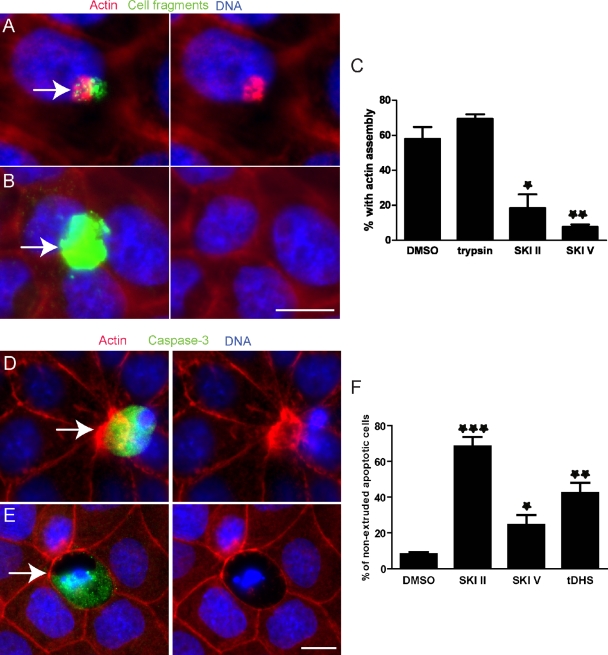
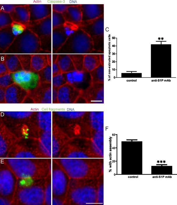
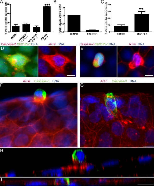
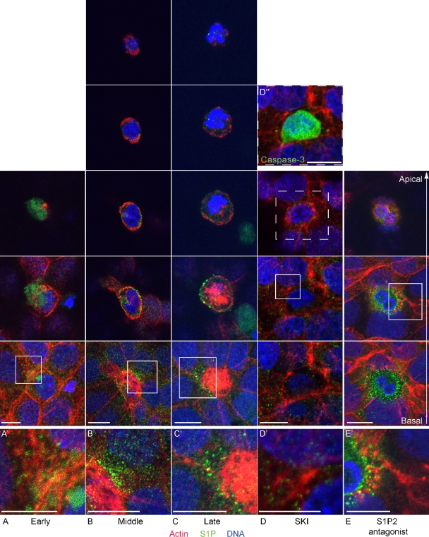
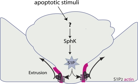
To maintain an intact barrier, epithelia eliminate dying cells by extrusion. During extrusion, a cell destined for apoptosis signals its neighboring cells to form and contract a ring of actin and myosin, which squeezes the dying cell out of the epithelium. Here, we demonstrate that the signal produced by dying cells to initiate this process is sphingosine-1-phosphate (S1P). Decreasing S1P synthesis by inhibiting sphingosine kinase activity or by blocking extracellular S1P access to its receptor prevented apoptotic cell extrusion. Extracellular S1P activates extrusion by binding the S1P(2) receptor in the cells neighboring a dying cell, as S1P(2) knockdown in these cells or its loss in a zebrafish mutant disrupted cell extrusion. Because live cells can also be extruded, we predict that this S1P pathway may also be important for driving delamination of stem cells during differentiation or invasion of cancer cells.


Figures

Figure 1.
Figure 1.
Inhibitors of SphKs block actin assembly and apoptotic cell extrusion. (A and B) Alexa Fluor 488–labeled cell fragments (green) prepared from MDCK cells pretreated with DMSO (A) or SKI II (B) were added to an intact MDCK monolayer. Arrows point to added cell fragments. (C) The percentage of cell fragments causing actin assembly from three independent experiments; n = 100 cell fragments per experiment and error bars are standard deviations (SDs). *, P < 0.05; **, P < 0.01. (D and E) Extrusion in an MDCK monolayer in the presence of DMSO (D) or SKI II (E). Arrows point to active caspase-3–positive dying cells in each case. (F) Quantification of nonextruded active caspase-3–positive apoptotic cells with DMSO or SphK inhibitor treatment from three independent experiments; n = 100, error bars = SDs. ***, P < 0.001. Bars, 10 µm.
Figure 2.
Figure 2.
An inhibitory anti-S1P mAb blocks apoptotic cell extrusion. (A and B) Extrusion in an MDCK monolayer treated with short-wave UV to induce apoptosis in the presence of a mouse IgG isotype control (A) or 10 µg/ml anti-S1P mAb (B). (C) Quantification of nonextruded apoptotic cells from three independent experiments; n = 100 active caspase-3–positive cells where error bars are SDs; **, P < 0.01. (D and E) Alexa Fluor 488–labeled cell fragments (green) prepared from MDCK cells were added to an intact MDCK monolayer in the presence of a mouse IgG isotype control (D) or 10 µg/ml anti-S1P mAb (E). (F) The percentage of cell fragments causing actin assembly from three independent experiments; n = 100 cell fragments per experiment and error bars = SDs. ***, P < 0.001. Bars, 10 µm.
Figure 3.
Figure 3.
Apoptotic cell extrusion requires the S1P2 receptor. (A) HBE cells induced to undergo apoptosis with UV in the presence of DMSO or the indicated S1P receptor antagonists. (B) qRT-PCR confirms shRNA-mediated knockdown of S1P2 in HBE cells. (C) Quantification of nonextruded apoptotic cells in HBE monolayers expressing control or S1P2-specific shRNA after UV treatment. (D and E) A dying HBE cell is not extruded by S1P2-silenced cells (D, green), but is extruded successfully by normal surrounding cells (E). When S1P2 shRNA is only in the dying cell (E), it extrudes and is in a higher plane than the actin ring below it, but is in the same plane when the surrounding cells are knocked down for S1P2 (D). Projections of extruding and nonextruding apoptotic cells from WT (F) or mil (G) zebrafish larvae, respectively. (H and I) Cross sections (XZs) of an apoptotic extruding cell (H) and a nonextruding cell (I) from WT (H) and mil zebrafish larvae (I), respectively. For all bar graphs, each bar represents the average percentage of nonextruded apoptotic cells to total apoptotic cells with each treatment from three independent experiments; n = 100 dying cells per experiment, error bars = SDs. **, P < 0.01; ***, P < 0.001. Bars, 10 µm.
Figure 4.
Figure 4.
Apoptotic cells produce and transmit S1P during extrusion. (A–C) Confocal fluorescence images during early (A), middle (B), and late (C) stages of extrusion of apoptotic cells from an HBE monolayer. (D and E) Confocal fluorescence images of blocked apoptotic cell extrusion by SKI II (D) or the S1P2 antagonist JTE-013 (E). Each experimental sample was visualized with five (B and C) or three (A, D, and E) consecutive 3D projections (comprising 2-µm thickness each), as necessary to span the full distance from the most basal to the most apical section (second-to-bottom and top images, respectively). Note that total cell height under the different conditions varies: during early extrusion (A) and when extrusion is blocked with SKI II and JTE-013 (D and E), the dying cell is not squeezed out of the epithelium and therefore does not inhabit as great an apical-to-basal distance as when the dying cell is extruding (B and C). A, B, C, and E were obtained using a confocal microscope (TCS SP5; Leica), whereas D was taken using an inverted microscope (Eclipse TE300; Nikon) converted for spinning disc confocal microscopy. A′–E′ represent zoomed-in region (square) from each montage. (D′′) Inset denoting that the unextruded cell in D is apoptotic. Bars, 10 µm.
Figure 5.
Figure 5.
Model for how S1P triggers extrusion. S1P is produced in the dying cell and exported to neighboring cells. S1P binds to S1P2 and triggers formation and contraction of an actin-and-myosin II extruding ring to squeeze the dying cell out.

References

    1. Brinkmann V., Davis M.D., Heise C.E., Albert R., Cottens S., Hof R., Bruns C., Prieschl E., Baumruker T., Hiestand P., et al. 2002. The immune modulator FTY720 targets sphingosine 1-phosphate receptors. J. Biol. Chem. 277:21453–21457 10.1074/jbc.C200176200 - DOI - PubMed
    1. Davis M.D., Clemens J.J., Macdonald T.L., Lynch K.R. 2005. Sphingosine 1-phosphate analogs as receptor antagonists. J. Biol. Chem. 280:9833–9841 10.1074/jbc.M412356200 - DOI - PubMed
    1. Estrada R., Zeng Q., Lu H., Sarojini H., Lee J.F., Mathis S.P., Sanchez T., Wang E., Kontos C.D., Lin C.Y., et al. 2008. Up-regulating sphingosine 1-phosphate receptor-2 signaling impairs chemotactic, wound-healing, and morphogenetic responses in senescent endothelial cells. J. Biol. Chem. 283:30363–30375 10.1074/jbc.M804392200 - DOI - PMC - PubMed
    1. French K.J., Schrecengost R.S., Lee B.D., Zhuang Y., Smith S.N., Eberly J.L., Yun J.K., Smith C.D. 2003. Discovery and evaluation of inhibitors of human sphingosine kinase. Cancer Res. 63:5962–5969 - PubMed
    1. Gibson M.C., Perrimon N. 2005. Extrusion and death of DPP/BMP-compromised epithelial cells in the developing Drosophila wing. Science. 307:1785–1789 10.1126/science.1104751 - DOI - PubMed

Publication types

MeSH terms