. 2011 Jun 15;186(12):7110-9.
doi: 10.4049/jimmunol.1100001. Epub 2011 May 9.

Lung neutrophils facilitate activation of naive antigen-specific CD4+ T cells during Mycobacterium tuberculosis infection

Affiliations

Lung neutrophils facilitate activation of naive antigen-specific CD4+ T cells during Mycobacterium tuberculosis infection

Robert Blomgran et al. J Immunol. .

Abstract

Initiation of the adaptive immune response to Mycobacterium tuberculosis occurs in the lung-draining mediastinal lymph node and requires transport of M. tuberculosis by migratory dendritic cells (DCs) to the local lymph node. The previously published observations that 1) neutrophils are a transiently prominent population of M. tuberculosis-infected cells in the lungs early in infection and 2) that the peak of infected neutrophils immediately precedes the peak of infected DCs in the lungs prompted us to characterize the role of neutrophils in the initiation of adaptive immune responses to M. tuberculosis. We found that, although depletion of neutrophils in vivo increased the frequency of M. tuberculosis-infected DCs in the lungs, it decreased trafficking of DCs to the mediastinal lymph node. This resulted in delayed activation (CD69 expression) and proliferation of naive M. tuberculosis Ag85B-specific CD4 T cells in the mediastinal lymph node. To further characterize the role of neutrophils in DC migration, we used a Transwell chemotaxis system and found that DCs that were directly infected by M. tuberculosis migrated poorly in response to CCL19, an agonist for the chemokine receptor CCR7. In contrast, DCs that had acquired M. tuberculosis through uptake of infected neutrophils exhibited unimpaired migration. These results revealed a mechanism wherein neutrophils promote adaptive immune responses to M. tuberculosis by delivering M. tuberculosis to DCs in a form that makes DCs more effective initiators of naive CD4 T cell activation. These observations provide insight into a mechanism for neutrophils to facilitate initiation of adaptive immune responses in tuberculosis.


Figures
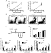
Figure 1
Figure 1
Myeloid cell and bacterial populations in the lungs of neutrophil depleted and control mice. (A) Neutrophil accumulation in the BAL and lung parenchyma during the early stages of M. tuberculosis infection. (B) Representative flow plots of lung neutrophils at day 17 post infection (when lung neutrophils are abundant), in mice treated with isotype or 1A8 three days earlier. Shown is the gating strategy routinely used to identify neutrophils (PMN) (Gr-1hi/CD11bhi); Gr-1/Ly6C staining was performed to evaluate and confirm the validity of the Gr-1/CD11b gating. Neutrophils were additionally CD11clo/neg (not shown). (C) Experimental scheme for analysis of effects of neutrophil depletion in M. tuberculosis-infected mice. Mice were infected with ~100 CFU/mouse. (D) Lung bacterial burdens in neutrophil-depleted and control mice. No statistically significant differences were observed. (E) Effects of neutrophil depletion on myeloid cell populations in the lungs on days 14, 17, and 21 postinfection. Data are mean ± SD of five mice per group and time point. The results shown in panels D and E are representative data of three separate experiments. PMN, neutrophils; AM, alveolar macrophages; mDC, myeloid dendritic cells; RM, recruited macrophages; Mo, monocytes. *, p < 0.05; **, p < 0.01; ***, p < 0.001.
Figure 2
Figure 2
Neutrophil depletion redistributes M. tuberculosis to other myeloid cells in the lungs. C57BL/6 mice aerosol infected with a low dose of GFP-expressing H37Rv were treated with 1A8 or isotype control antibody at day 16 post infection and analyzed at day 19 (A) or day 21 (B). No difference in the bacterial burden in lung or MDLN between groups was detected. Data are mean ± SD of five mice per group and time point. PMN, neutrophils; AM, alveolar macrophages; mDC, myeloid dendritic cells; RM, recruited macrophages; Mo, monocytes. **, p < 0.01.
Figure 3
Figure 3
Neutrophils accelerate activation of naïve M. tuberculosis Ag85B-specific CD4+ T cells in vivo. Mice were treated as shown in Figure 1C after adoptive transfer of naïve P25TCR-Tg CD4+ T cells on the day preceding infection. (A) Representative CFSE-dilution profiles of P25TCR-Tg CD4+ T cells in MDLN on days 14, 15, and 17 postinfection. (B) P25TCR-Tg cell activation as reflected by CD69 expression; gating used is shown in panel A. (C) Comparison of P25TCR-Tg CD4+ T cell proliferation in neutrophil-depleted and control mice; gating used is shown in panel A. Data in panels B and C are mean ± SD of five mice per group and time point. Data are representative of two separate experiments. **, p < 0.01.
Figure 4
Figure 4
Neutrophils accelerate trafficking of M. tuberculosis and dendritic cells from the lungs to the mediastinal lymph node. Mice were treated as shown in Figure 1C. (A) M. tuberculosis CFU in MDLN homogenates at the indicated time-points. (B) CD11bhigh/CD11chigh myeloid dendritic cells on days 14 and 15 postinfection. Data are mean ± SD of five mice per group and time point, representating data from two (A) or one (B) separate experiments. Data in B were analyzed using unpaired one-tailed Student t test. *, p < 0.05.
Figure 5
Figure 5
M. tuberculosis -infected neutrophils provide antigen to dendritic cells for presentation to CD4+ T cells. (A) BMDCs were directly infected with M. tuberculosis (Mtb; MOI=5) or incubated with an equal number of neutrophils that were previously infected with M. tuberculosis (MOI=5) for 19 hours. Thereafter, BMDCs were incubated with P25TCR-Tg Th1 effector CD4+ T cells, and release of IFNγ into supernatants was analyzed after 6, 12, 24 and 36 hours. (B) BMDCs were directly infected with M. tuberculosis at the MOI shown or incubated with an equal number of M. tuberculosis-infected neutrophils (previously infected at the MOI shown), then incubated with P25TCR-Tg Th1 effector CD4+ T cells, and release of IFNγ into supernatants was analyzed after 12 hours. (C) CFSE-labeled naïve P25TCR-Tg CD4+ T cells were added to BMDC that were either directly infected or after incubation with infected neutrophils as described for panels 5A and B, and T-cell proliferation was analyzed at 72 hours. Mean values of cells that had undergone at least one cycle of proliferation are depicted in histograms. Data in A–C are representative of two separate experiments performed in triplicate. Un = undetected.
Figure 6
Figure 6
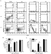
M. tuberculosis inhibits DC migration when DCs acquire bacteria directly, but not when they acquire bacteria by ingesting infected neutrophils. Before migration assays, DCs were directly infected with GFP-M. tuberculosis (MOI=5), or incubated with an equal number of either uninfected or infected (MOI=5) CMTMR-labeled neutrophils for 19 hours. To distinguish migration of dendritic cells that contained bacteria, neutrophils, both, or neither, input cells and cells in the lower well of the Transwell chamber after migration were stained with antibodies to CD11c and Ly6G, counted, and analyzed by flow cytometry. (A) Shows the gating strategy used to exclude extracellular neutrophils (Ly6G+; top panel), and R1–R4 (bottom panel) distinguish DCs that contain M. tuberculosis (GFP+), infected neutrophils (CMTMR+/GFP+), or uninfected neutrophils (CMTMR+/GFP−) (BMDCs used as input cells are displayed). (B) BMDCs were allowed to migrate 3 hours in the presence of CCL19. (C) CCR7 expression on uninfected DCs, directly-infected DCs, and DCs that acquired M. tuberculosis by ingesting infected neutrophils. (D) In vivo expanded spleen DCs (Flt3L-DC) did not ingest neutrophils, but could be infected by M. tuberculosis, restricting the evaluation of Flt3L-DC migration to cells incubated with M. tuberculosis (containing bacteria or not) and untreated DCs. In addition to GFP+ DCs, GFP− DCs from the M. tuberculosis stimulated group were also analyzed. Unstimulated CCR7−/− Flt3L-DCs were used to determine baseline random migration. Data are expressed as mean ± SD percent of the input cells that migrated to the lower chamber after 1 hour. Each condition was assayed in triplicate; data are representative of two (B and C) and three (D) independent experiments. Mtb, M. tuberculosis; infPMN, M. tuberculosis-infected neutrophils; uninfPMN, uninfected neutrophils. *, p < 0.05; **, p < 0.01.
Figure 7
Figure 7
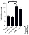
M. tuberculosis -infected neutrophils release one or more chemoattractants for DCs. Particle free and sterile filtered medium from uninfected neutrophils (o/n PMN-sup) or M. tuberculosis infected neutrophils (o/n infPMN-sup) were used as chemotactic stimuli in the lower chamber of Transwell chambers. Data are expressed as mean ± SD percent of the input cells that migrated to the lower chamber after 3 hours. Each condition was performed in triplicate; data are representative of three independent experiments. **, p < 0.01.

References

    1. Meena LS, Rajni Survival mechanisms of pathogenic Mycobacterium tuberculosis H37Rv. FEBS J. 2010;277:2416–2427. - PubMed
    1. Mogues T, Goodrich ME, Ryan L, LaCourse R, North RJ. The relative importance of T cell subsets in immunity and immunopathology of airborne Mycobacterium tuberculosis infection in mice. J Exp Med. 2001;193:271–280. - PMC - PubMed
    1. Gallegos AM, Pamer EG, Glickman MS. Delayed protection by ESAT-6-specific effector CD4+ T cells after airborne M. tuberculosis infection. J Exp Med. 2008;205:2359–2368. - PMC - PubMed
    1. Hoang TT, Nansen A, Roy S, Billeskov R, Aagaard C, Elvang T, Dietrich J, Andersen P. Distinct differences in the expansion and phenotype of TB10.4 specific CD8 and CD4 T cells after infection with Mycobacterium tuberculosis. PLoS One. 2009;4:e5928. - PMC - PubMed
    1. Woodworth JS, Behar SM. Mycobacterium tuberculosis-specific CD8+ T cells and their role in immunity. Crit Rev Immunol. 2006;26:317–352. - PMC - PubMed

Publication types

Substances