

Tracking brain states under general anesthesia by using global coherence analysis
- PMID: 21555565
- PMCID: PMC3102391
- DOI: 10.1073/pnas.1017041108
Tracking brain states under general anesthesia by using global coherence analysis
Abstract
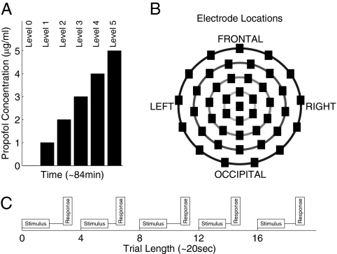
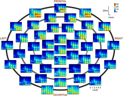
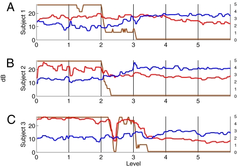
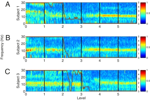
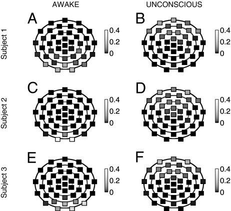
Time and frequency domain analyses of scalp EEG recordings are widely used to track changes in brain states under general anesthesia. Although these analyses have suggested that different spatial patterns are associated with changes in the state of general anesthesia, the extent to which these patterns are spatially coordinated has not been systematically characterized. Global coherence, the ratio of the largest eigenvalue to the sum of the eigenvalues of the cross-spectral matrix at a given frequency and time, has been used to analyze the spatiotemporal dynamics of multivariate time-series. Using 64-lead EEG recorded from human subjects receiving computer-controlled infusions of the anesthetic propofol, we used surface Laplacian referencing combined with spectral and global coherence analyses to track the spatiotemporal dynamics of the brain's anesthetic state. During unconsciousness the spectrograms in the frontal leads showed increasing α (8-12 Hz) and δ power (0-4 Hz) and in the occipital leads δ power greater than α power. The global coherence detected strong coordinated α activity in the occipital leads in the awake state that shifted to the frontal leads during unconsciousness. It revealed a lack of coordinated δ activity during both the awake and unconscious states. Although strong frontal power during general anesthesia-induced unconsciousness--termed anteriorization--is well known, its possible association with strong α range global coherence suggests highly coordinated spatial activity. Our findings suggest that combined spectral and global coherence analyses may offer a new approach to tracking brain states under general anesthesia.
Conflict of interest statement
The authors declare no conflict of interest.
Figures

References
-
- Gibbs F, Gibbs E, Lennox W. Effect on the electroencephalogram of certain drugs which influence nervous activity. Arch Intern Med. 1937;60:154–166.
-
- Kiersey DK, Bickford RG, Faulconer A., Jr Electro-encephalographic patterns produced by thiopental sodium during surgical operations; description and classification. Br J Anaesth. 1951;23:141–152. - PubMed
-
- Tinker JH, Sharbrough FW, Michenfelder JD. Anterior shift of the dominant EEG rhytham during anesthesia in the Java monkey: Correlation with anesthetic potency. Anesthesiology. 1977;46:252–259. - PubMed
-
- Kearse LA, Jr, Manberg P, Chamoun N, deBros F, Zaslavsky A. Bispectral analysis of the electroencephalogram correlates with patient movement to skin incision during propofol/nitrous oxide anesthesia. Anesthesiology. 1994;81:1365–1370. - PubMed
-
- Billard V, Gambus PL, Chamoun N, Stanski DR, Shafer SL. A comparison of spectral edge, delta power, and bispectral index as EEG measures of alfentanil, propofol, and midazolam drug effect. Clin Pharmacol Ther. 1997;61:45–58. - PubMed
Publication types
MeSH terms
Grants and funding
LinkOut - more resources
Full Text Sources
Other Literature Sources
Medical
