. 2011 May 24;108(21):8885-90.
doi: 10.1073/pnas.1105564108. Epub 2011 May 9.

Dual and opposing roles of the unfolded protein response regulated by IRE1alpha and XBP1 in proinsulin processing and insulin secretion

Affiliations

Dual and opposing roles of the unfolded protein response regulated by IRE1alpha and XBP1 in proinsulin processing and insulin secretion

Ann-Hwee Lee et al. Proc Natl Acad Sci U S A. .

Abstract

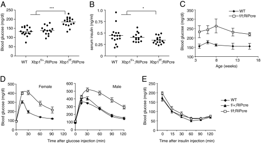
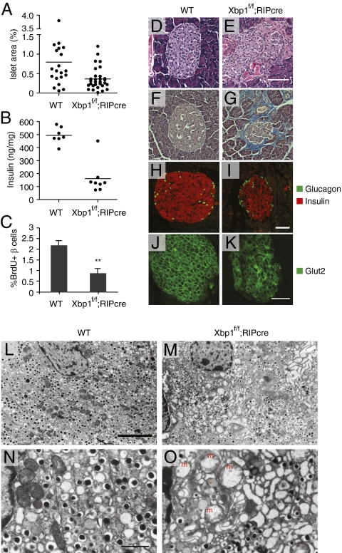
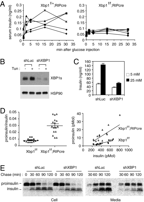
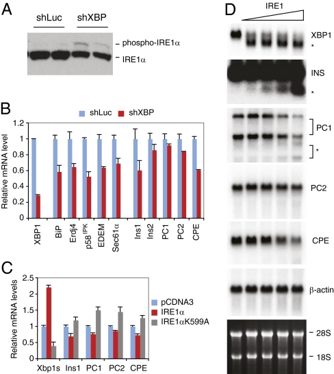
As a key regulator of the unfolded protein response, the transcription factor XBP1 activates genes in protein secretory pathways and is required for the development of certain secretory cells. To elucidate the function of XBP1 in pancreatic β-cells, we generated β-cell-specific XBP1 mutant mice. Xbp1(f/f);RIP-cre mice displayed modest hyperglycemia and glucose intolerance resulting from decreased insulin secretion from β-cells. Ablation of XBP1 markedly decreased the number of insulin granules in β-cells, impaired proinsulin processing, increased the serum proinsulin:insulin ratio, blunted glucose-stimulated insulin secretion, and inhibited cell proliferation. Notably, XBP1 deficiency not only compromised the endoplasmic reticulum stress response in β-cells but also caused constitutive hyperactivation of its upstream activator, IRE1α, which could degrade a subset of mRNAs encoding proinsulin-processing enzymes. Hence, the combined effects of XBP1 deficiency on the canonical unfolded protein response and its negative feedback activation of IRE1α caused β-cell dysfunction in XBP1 mutant mice. These results demonstrate that IRE1α has dual and opposing roles in β-cells, and that a precisely regulated feedback circuit involving IRE1α and its product XBP1s is required to achieve optimal insulin secretion and glucose control.


Conflict of interest statement

Conflict of interest statement: L.H.G. is a member of the board of directors of and holds equity in the Bristol Myers Squibb Corporation.

Figures

Fig. 1.
Fig. 1.
β-cell-specific deletion of Xbp1 causes hyperglycemia and glucose intolerance in mice. (A) Blood glucose and (B) serum insulin levels were measured after fasting mice for 6 h. Each dot represents an individual male mouse (7-wk-old). (C) Fed-state blood glucose levels of WT and β-cell-specific Xbp1 knockout (Xbp1f/f;RIP-cre) mice were measured along the time course. n = 6–8 male mice per group. Values represent means ± SEM. (D) GTTs were performed on 7- to 8-wk-old mice. n = 6–8 mice per group. (E) ITT was performed on 16-wk-old male mice. n = 5–7 mice per group. *P < 0.05; ***P < 0.001.
Fig. 2.
Fig. 2.
Histopathological analysis of islets of β-cell-specific Xbp1 KO mice. (A) Islet area relative to total pancreas. One random pancreas section per mouse was examined. Each dot represents an individual mouse of the indicated genotype of 4–6 mo of age. (B) Pancreatic insulin contents of 12- to 16-wk-old male mice. (C) BrdU-positive β-cells were counted after BrdU/insulin double staining. n = 4–5 mice per group. **P < 0.01. Pancreatic sections from WT and Xbp1f/f;RIP-cre mice were stained with (D and E) hematoxylin and eosin, (F and G) trichrome blue, (H and I) insulin and glucagon, and (J and K) Glut2 antibodies followed by fluorescence-conjugated secondary antibodies. (Scale bars, 100 μm.) (LO) Islet sections were examined by TEM. [Scale bar, 5 μm (L and M); 1 μm (N and O).] The asterisk indicates an electron lucent granule. m, swollen mitochondria.
Fig. 3.
Fig. 3.
Impaired insulin secretion and proinsulin maturation in XBP1-deficient β-cells. (A) Glucose-stimulated insulin secretion assays. Serum insulin levels were measured in control heterozygous Xbp1f/+;RIP-cre and Xbp1f/f;RIP-cre mice at indicated time points after a bolus glucose injection. Each line represents an individual mouse. (B) Min6 cells stably expressing shRNAs targeting control luciferase or XBP1 mRNAs were tested for XBP1s expression by Western blot. (C) Cells were pretreated with 2.5 mM glucose for 2 h and then cultured in 5 mM or 25 mM glucose media for 2 h. Culture media were collected to measure insulin content by ELISA. (D) Serum proinsulin levels relative to total insulin in WT and Xbp1f/f;RIP-cre mice were determined. Data from both males and females were combined, because sex difference was not significant. The graphs display proinsulin:insulin ratio (Left) or proinsulin and insulin concentrations (pmol per liter) (Right). Each dot represents an individual mouse (4- to 6-mo-old). (E) Min6 cells expressing control or XBP1 shRNA were pulse-labeled for 30 min with [35S]Met/Cys and then cultured in chase media for the indicated time. Cells and culture supernatants were harvested for immunoprecipitation of radiolabeled insulin and proinsulin, which were revealed by SDS/PAGE followed by autoradiography.
Fig. 4.
Fig. 4.
Feedback activation of IRE1α by XBP1 deficiency, and IRE1α-mediated mRNA cleavage. (A) Western blot and (B) Quantitative RT-PCR analysis of Min6 cells expressing luciferase or XBP1 shRNAs. (C) 293T cells were cotransfected with EGFP, Ins1, PC1, PC2, and CPE plasmids together with WT or K599A mutant IRE1α. Cells were harvested 24 h after transfection for quantitative RT-PCR analysis. EGFP mRNA levels were used for normalization. Values represent fold changes relative to vector controls. (D) Total Min6 RNA was incubated with increasing amounts of recombinant IRE1α and then separated on an agarose gel. Northern blot was performed using the indicated 32P-labeled probes. The asterisks indicate cleavage products.

References

    1. Schröder M, Kaufman RJ. The mammalian unfolded protein response. Annu Rev Biochem. 2005;74:739–789. - PubMed
    1. Lee AH, Glimcher LH. Intersection of the unfolded protein response and hepatic lipid metabolism. Cell Mol Life Sci. 2009;66:2835–2850. - PMC - PubMed
    1. Ron D, Walter P. Signal integration in the endoplasmic reticulum unfolded protein response. Nat Rev Mol Cell Biol. 2007;8:519–529. - PubMed
    1. Kaufman RJ. Stress signaling from the lumen of the endoplasmic reticulum: Coordination of gene transcriptional and translational controls. Genes Dev. 1999;13:1211–1233. - PubMed
    1. Lee AH, Iwakoshi NN, Glimcher LH. XBP-1 regulates a subset of endoplasmic reticulum resident chaperone genes in the unfolded protein response. Mol Cell Biol. 2003;23:7448–7459. - PMC - PubMed

Publication types