. 2011 May 24;108(21):E128-35.
doi: 10.1073/pnas.1101964108. Epub 2011 May 9.

Hypomorphic Notch 3 alleles link Notch signaling to ischemic cerebral small-vessel disease

Affiliations

Hypomorphic Notch 3 alleles link Notch signaling to ischemic cerebral small-vessel disease

Joseph F Arboleda-Velasquez et al. Proc Natl Acad Sci U S A. .

Abstract

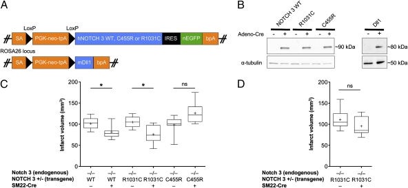
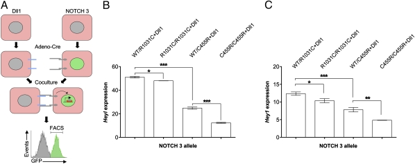
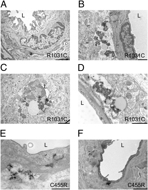
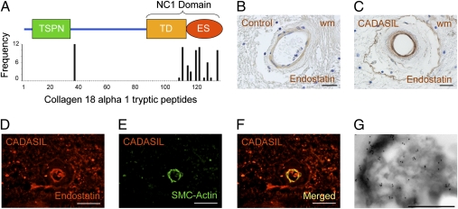
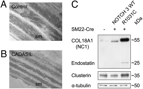
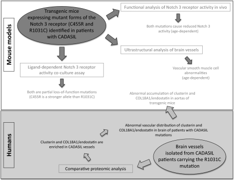
The most common monogenic cause of small-vessel disease leading to ischemic stroke and vascular dementia is the neurodegenerative syndrome cerebral autosomal-dominant arteriopathy with subcortical infarcts and leukoencephalopathy (CADASIL), which is associated with mutations in the Notch 3 receptor. CADASIL pathology is characterized by vascular smooth muscle cell degeneration and accumulation of diagnostic granular osmiophilic material (GOM) in vessels. The functional nature of the Notch 3 mutations causing CADASIL and their mechanistic connection to small-vessel disease and GOM accumulation remain enigmatic. To gain insight into how Notch 3 function is linked to CADASIL pathophysiology, we studied two phenotypically distinct mutations, C455R and R1031C, respectively associated with early and late onset of stroke, by using hemodynamic analyses in transgenic mouse models, receptor activity assays in cell culture, and proteomic examination of postmortem human tissue. We demonstrate that the C455R and R1031C mutations define different hypomorphic activity states of Notch 3, a property linked to ischemic stroke susceptibility in mouse models we generated. Importantly, these mice develop osmiophilic deposits and other age-dependent phenotypes that parallel remarkably the human condition. Proteomic analysis of human brain vessels, carrying the same CADASIL mutations, identified clusterin and collagen 18 α1/endostatin as GOM components. Our findings link loss of Notch signaling with ischemic cerebral small-vessel disease, a prevalent human condition. We determine that CADASIL pathophysiology is associated with hypomorphic Notch 3 function in vascular smooth muscle cells and implicate the accumulation of clusterin and collagen 18 α1/endostatin in brain vessel pathology.


Conflict of interest statement

The authors declare no conflict of interest.

Figures

Fig. 1.
Fig. 1.
CADASIL mutations in Notch 3 reflect hypomorphic receptor activity in vivo. (A) Transgenic mouse models for in vivo studies of Notch signaling. Schematic representation of the targeting vectors used to generate conditional knock-in mice expressing human Notch 3 WT (hNOTCH3 WT), or the C455R or R1031C mutant receptors, or the mouse Dll1 ligand (also see ref. 16). Each construct contains genomic sequences allowing for homologous recombination into the ROSA locus; an adenovirus splice acceptor (SA); a PGK-neo-tpA “stop” cassette flanked by loxP sites (black triangles) allowing for Cre-mediated excision; the transgene; and the bovine growth hormone polyadenylation sequence (bpA). The NOTCH 3 WT and mutant transgenes are followed by an internal ribosomal entry site (IRES) and nuclear EGFP (nEGFP). (B) Western blot analysis of protein cell lysates shows conditional expression of Notch 3 receptors (S1-cleaved N fragment containing the transmembrane and intracellular domains, ∼90 kDa, in aorta-derived SMC cultures) and of Dll1 ligand (∼80 KDa, in MEFs), upon infection with adeno-Cre. The Notch 3 WT and mutant receptors are expressed at similar levels allowing for comparative studies. (C and D) Functional analysis of receptor activity in vivo presented as box-and-whisker plots. Cerebral infarcts were produced by MCA occlusion (1 h) followed by reperfusion in adult Notch 3 KO male mice expressing the different NOTCH 3 transgenes as indicated. Infarct volumes are calculated by integrating the infarct area in each brain section, using the indirect method to correct for edema (Methods). (C) In 3- to 6-mo-old mice, expression of WT or R1031C mutant receptors in vSMCs, using the SM22-Cre driver, rescues the ischemia susceptibility phenotype of Notch 3 KO animals, whereas expression of C455R mutant receptors does not. (D) In 1-y-old mice, expression of R1031C mutant receptors in vSMCs no longer rescues the ischemia susceptibility phenotype. The line bisecting the box in the plot is the median value, the cross is the mean, the upper and lower limits of the box are the 25th and 75th percentile values; the whiskers extend to the lowest and highest values. At least five animals were analyzed in each experimental group (Methods).
Fig. 2.
Fig. 2.
CADASIL mutations reflect hypomorphic Notch 3 receptor activity in vitro. (A) Schematic of the ligand-dependent Notch receptor activity coculture assay. MEFs isolated from mice were induced to express Notch 3 WT or mutant receptors or the Dll1 ligand using adeno-Cre infection and then cocultured for 48 h. Receptor-expressing cells were isolated by FACS. (B and C) Expression of Heyl (B) and Hey1 (C) in receptor-expressing cells assessed by qPCR. MEFs carrying two copies of the transgenes in homozygous (R1031C/R1031C and C455R/C455R) or heterozygous (WT/R1031C and WT/C455R) combinations were analyzed. The expression of Heyl and Hey1 was significantly lower in MEFs expressing homozygous combinations of the CADASIL alleles than their heterozygous counterparts. These results define an allelic series with the C455R mutation representing the stronger loss-of-Notch-function allele. Error bars indicate SDs. Data are representative of three independent experiments. Note that the activation of Heyl expression in this Notch signaling assay is far more robust than that of Hey1 across experiments. Statistically significant differences: *P < 0.05, **P < 0.01, and ***P < 0.005.
Fig. 3.
Fig. 3.
CADASIL transgenic mice develop vSMC abnormalities. (AD) Representative electron micrographs from 12- to 14-mo-old (AC) (n = 2) or 19- to 21-mo-old (D) (n = 2) animals carrying the human R1031C mutation in a mouse Notch 3 WT background. Morphological abnormalities were absent in animals carrying the R1031C mutation at 6 mo of age (n = 2). (E and F) Representative EM images from 6-mo-old animals carrying the C455R mutation in a Notch 3 KO background (n = 2). (A) Overview of a vessel with abundant elastin and an electron-dense granular deposit (arrow). (B) Accumulation of extracellular granular material (arrow) and cytoplasmic inclusions (asterisk). (C) An almost occluded vessel with accumulations of granular material. (D) Accumulation of granular material and inclusions (arrow). (E) Accumulation of electron-dense granular material (arrows). (F) Inclusions (asterisk) and electron-dense granular material (arrow). The brain tissue analyzed was compared with tissue from age-matched controls expressing the WT NOTCH 3 (n = 5) and from animals that lacked the SM22-Cre driver (n = 5) processed in parallel. L, lumen. (Scale bars: A, 2 μm; B and C, 1 μm; D and F, 2 μm; E, 500 nm.)
Fig. 4.
Fig. 4.
Molecular characterization of postmortem cerebral vessels from individuals with CADASIL. (A) Coronal MRI of the proband individual of the Colombian kindred with CADASIL carrying the R1031C mutation shows extensive bilateral leukoencephalopathy. (B) Photograph of postmortem coronal section of the left cerebral hemisphere at the level of the lateral geniculate nucleus shows loss of white matter, evident in the centrum semiovale (arrow). (C) Photomicrograph of deep subcortical white matter stained with Luxol fast blue (LFB)/PAS) shows marked hyalinization of the vessel walls, rare myelin sheaths (in blue), and PAS-positive deposits (arrow). (D) EM images of subcortical white matter of postmortem brains from individuals carrying the R1031C (Upper) and C455R (Lower) mutations show abundant electron-dense GOM deposits. (EH) Immunohistochemical staining shows abnormal accumulation of clusterin in brains from individuals carrying the R133C mutation (F and H) compared with age-matched controls (EG). Consistent with the severity of vessel pathology, clusterin staining is stronger in white matter (wm) than in gray matter (gm) arteries. (I and J) Clusterin is localized to the tunica media of the arteries but is excluded from the few remaining smooth muscle cells, labeled with α-actin (SMC-actin), in tissue from an individual with the R133C mutation. (K and L) Brain tissue from an individual with the C455R mutation was processed for immunogold labeling with an antibody against clusterin, which is detected in GOMs. (Scale bars: C and EI, 50 μm; D, 5 μm; K, 500 nm; L, 200 nm.)
Fig. 5.
Fig. 5.
Analysis of COL18A1/endostatin expression in cerebral vessels from individuals with CADASIL. (A) Schematic representation of COL18A1, isoform 2 (long; also see ref. 31). The N-terminal thrombospondin-like domain (TSPN) and the noncollagenous domain 1 (NC1) comprising the trimerization domain (TD) and the C-terminal endostatin domain (ES) are indicated. The graph shows the frequency of peptides along the length of COL18A1 detected by mass spectroscopy of arterial rings microdissected from postmortem brain sections of two individuals with the R1031C mutation. (B and C) Immunohistochemistry (IHC) with COL18A1/endostatin-specific antibodies shows abnormal distribution of this protein in the white matter (wm) of an individual carrying the R133C mutation (C) compared with control (B). (DF) COL18A1/endostatin localizes primarily to the smooth muscle cell layer, labeled with α-actin (SMC-actin) in brain tissue from a R133C carrier. (G) Brain tissue from an individual with the R1031C (G) mutation was processed for immunogold labeling with an antibody against COL18A1/endostatin, which associates with GOMs. (Scale bars: B and C, 25 μm; D–F, 50 μm; G, 1 μm.)
Fig. 6.
Fig. 6.
Clusterin and COL18A1/endostatin are misregulated in transgenic mice expressing mutant Notch 3 receptors. (A and B) EM images of aortas from 12- to 14-mo-old animals expressing the R1031C mutation (B) show thinner layers of vSMCs compared with controls (A). (C) Western blot analysis of aortic protein extracts derived from 18-mo-old mice expressing WT Notch 3 or the R1031C mutation shows increased levels of clusterin (∼35 kDa) and endostatin (∼25 kDa) in mice expressing the mutant receptor. The endostatin antibody used for immunoblotting recognizes endostatin whether free (cleaved from COL18A1) or part of the COL18A1 molecule. The expression of an approximately 55-kDa protein, likely corresponding to the NC1 domain of COL18A1, is also significantly increased in the aorta of mutant animals compared with controls.
Fig. 1.
Fig. 1.
Experimental design and key findings. Transgenic mouse models conditionally expressing two mutant forms of the Notch 3 receptor identified in Colombian families afflicted with CADASIL, each defining a particular phenotype of the disease, were generated. They were used to examine the activity of Notch 3 receptors carrying the C455R or R1031C mutations by using functional assays in vivo, ultrastructural analysis, and a ligand-dependent assay of Notch 3 receptor activity in cell culture. Both mutations are loss-of-function mutations: the early-onset C455R mutation represents a more severe loss of Notch 3 receptor function than the late-onset R1031C mutation, and both result in age-dependent phenotypes in the transgenic animal models, including accumulation of deposits in brain vessels. In parallel, brain blood vessels were collected from postmortem tissue carrying the R1031C mutation and analyzed by using MS. The proteomic analysis identified several proteins enriched in CADASIL vessels, including clusterin and collagen 18 α1/endostatin. Both proteins showed abnormal distribution in brain vessels of patients with several CADASIL mutations and, moreover, their expression was misregulated in the transgenic animal models expressing the mutant Notch 3 receptors. These findings point to remarkable similarity between the human disease and the transgenic animal models characterized in this study.

References

    1. Thompson CS, Hakim AM. Living beyond our physiological means: Small vessel disease of the brain is an expression of a systemic failure in arteriolar function: A unifying hypothesis. Stroke. 2009;40:e322–e330. - PubMed
    1. Joutel A, et al. Notch3 mutations in CADASIL, a hereditary adult-onset condition causing stroke and dementia. Nature. 1996;383:707–710. - PubMed
    1. Gould DB, et al. Role of COL4A1 in small-vessel disease and hemorrhagic stroke. N Engl J Med. 2006;354:1489–1496. - PubMed
    1. Revesz T, et al. Cerebral amyloid angiopathies: a pathologic, biochemical, and genetic view. J Neuropathol Exp Neurol. 2003;62:885–898. - PubMed
    1. Hara K, et al. Association of HTRA1 mutations and familial ischemic cerebral small-vessel disease. N Engl J Med. 2009;360:1729–1739. - PubMed

Publication types

LinkOut - more resources