. 2011 May;129(5):649-52.
doi: 10.1001/archophthalmol.2011.84.

Two novel CHN1 mutations in 2 families with Duane retraction syndrome

Affiliations

Two novel CHN1 mutations in 2 families with Duane retraction syndrome

Wai-Man Chan et al. Arch Ophthalmol. 2011 May.

Abstract

Objective: To determine the genetic cause of Duane retraction syndrome (DRS) in 2 families segregating DRS as a dominant trait.

Methods: Members of 2 unrelated pedigrees were enrolled in a genetic study. Linkage analysis was performed on the CHN1 locus. Probands and family members were screened for CHN1 mutations.

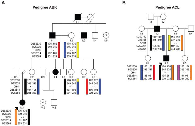
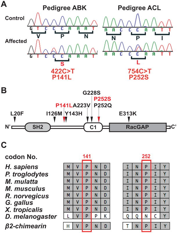
Results: The 6 affected individuals in the 2 pedigrees have DRS. Both pedigrees are consistent with linkage to the locus. Sequence analysis revealed 2 novel heterozygous missense CHN1 mutations, c.422C>T and c.754C>T, predicted to result in α2-chimaerin amino acid substitutions P141L and P252S, respectively.

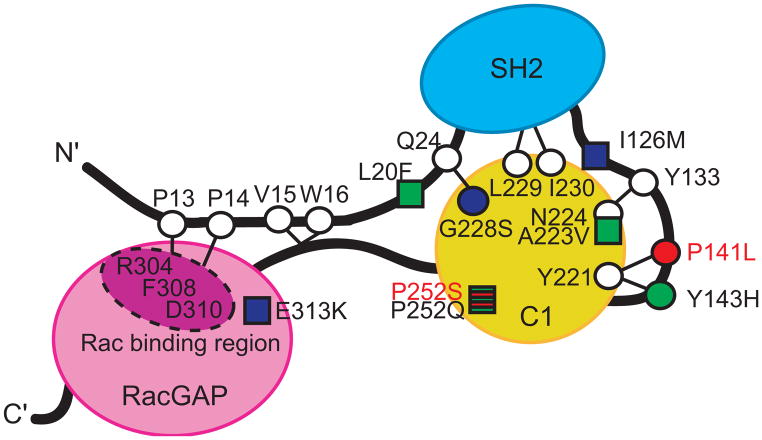
Conclusions: Genetic analysis of 2 pedigrees revealed 2 novel DRS mutations, bringing the number of DRS pedigrees known to harbor CHN1 from 7 to 9. Both mutations alter residues that participate in intramolecular interactions that stabilize the inactive, closed conformation of α2-chimaerin and, thus, are predicted to result in its hyperactivation. Moreover, amino acid residue P252 was previously reported to be altered to a different residue in a previously reported DRS pedigree; thus, this is the first report of 2 CHN1 mutations altering the same residue, further supporting a gain-of-function etiology.

Clinical relevance: Members of families segregating DRS as an autosomal dominant trait should be screened for mutations in the CHN1 gene, enhancing genetic counseling and permitting earlier diagnosis.


Figures

Figure 1
Figure 1. Schematic pedigrees and haplotype analysis of families segregating autosomal dominant DRS at the CHN1 locus
Pedigree members are denoted by circles (females) and squares (males) and by their generation and position. Black circles/squares indicate clinically affected individuals, and black arrowheads denote the probands. Haplotype analysis of (A) pedigree ABK and (B) pedigree ACL are shown schematically for markers surrounding the CHN1 gene. Genotyping data and schematic segregating haplotype bars for four chromosome 2 markers and CHN1 mutation status are shown below the symbol for each individual who participated in the study. Black bars indicate the affected haplotype that was passed from the affected grandfather in pedigree ABK or the affected father in pedigree ACL. For CHN1, (+) indicates presence of the mutation while (−) indicates its absence. For Pedigree ABK, the affected haplotype and CHN1 mutation are also inherited by two unaffected family members, III:2 and III:6.
Figure 2
Figure 2. Nucleotide sequence, amino acid position, and conservation of the CHN1 mutations
(A) Heterozygous CHN1 mutations in probands of pedigree ABK and ACL. Sequence chromatographs of the control individuals are normal (top row), while sequence chromatographs of the affected individual with DRS (bottom row) each reveals a heterozygous CHN1 mutation. The normal sequence and corresponding amino acid residues are indicated under each control sequence chromatograph (black), while the mutation and resulting amino acid substitution are denoted under each affected sequence (red). (B) Predicted α2-chimaerin protein structure. The amino acid residues altered by the two novel heterozygous mutations are depicted in red, with red arrows above the protein pointing to their predicted positions. The seven previously reported CHN1 mutations are indicated in black and their locations are indicated by black arrows above the protein. (C) Portions of the human CHN1 amino acid sequence that surround each substitution are aligned with homologous sequence in seven different species, followed by alignment with the paralogous β2-chimerin sequence at the bottom. Identical amino acid residues are highlighted in light gray. The residues altered by the two new mutations are boxed in red.
Figure 3
Figure 3. Schematic of α2-chimaerin structure in its closed conformation showing the predicted α2-chimaerin intramolecular interactions
The three domains of α2-chimaerin are depicted as follows: the N-terminal Src homology-2 (SH2) domain is depicted in blue, the C1 domain that binds to the second message signaling lipid diacylglycerol (DAG) is depicted in yellow; and the RacGAP domain that interacts with Rac and down-regulates its activity is depicted in pink. Linker regions are depicted as black lines. Specific amino acid residues are highlighted as circles or squares, with circles representing the positions of amino acids predicted to be involved in intramolecular interactions that stabilize the closed conformation of α2-chimaerin based on homology with β2-chimaerin. The seven previously reported mutations alter amino acid residues that are represented by circles or squares filled with green or blue; those filled with green were previously demonstrated to enhance translocation of α2-chimaerin to the membrane when mutated, while those filled with blue did not. The circle filled with red and the square striped red and green represent the residues altered by the new novel mutations: P252S alters the same residue as P252Q (thus the residue is striped), while P14lL alters a residue predicted to interact with Y221. Thus, both residues are anticipated to destablize the closed conformation of α2-chimaerin and result in its pathological hyperactivation. Figure adapted with permission from Miyake et al.

References

    1. Engle EC. The genetic basis of complex strabismus. Pediatr Res. 2006 Mar;59(3):343–348. - PubMed
    1. Kirkham T. Inheritance of Duane’s syndrome. Br J Ophthalmol. 1970;54:323–329. - PMC - PubMed
    1. DeRespinis PA, Caputo AR, Wagner RS, Guo S. Duane’s retraction syndrome. Surv Ophthalmol. 1993;38(3):258–288. - PubMed
    1. Hotchkiss MG, Miller NR, Clark AW, Green WG. Bilateral Duane’s retraction syndrome: A clinical-pathological case report. Arch Ophthalmol. 1980 May;98:870–874. - PubMed
    1. Miller NR, Kiel SM, Green WR, Clark AW. Unilateral Duane’s retraction syndrome (type 1) Arch Ophthalmol. 1982 Sep;100(9):1468–1472. - PubMed

Publication types