

Test-retest reliability of memory task functional magnetic resonance imaging in Alzheimer disease clinical trials
- PMID: 21555634
- PMCID: PMC3291175
- DOI: 10.1001/archneurol.2011.94
Test-retest reliability of memory task functional magnetic resonance imaging in Alzheimer disease clinical trials
Abstract
Objective: To examine the feasibility and test-retest reliability of encoding-task functional magnetic resonance imaging (fMRI) in mild Alzheimer disease (AD).
Design: Randomized, double-blind, placebo-controlled study.
Setting: Memory clinical trials unit.
Participants: We studied 12 patients with mild AD (mean [SEM] Mini-Mental State Examination score, 24.0 [0.7]; mean Clinical Dementia Rating score, 1.0) who had been taking donepezil hydrochloride for more than 6 months from the placebo arm of a larger 24-week study (n = 24, 4 scans on weeks 0, 6, 12, and 24, respectively).
Interventions: Placebo and 3 face-name, paired-associate encoding, block-design blood oxygenation level-dependent fMRI scans in 12 weeks.
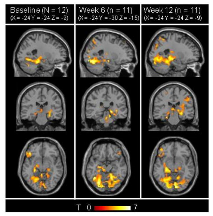
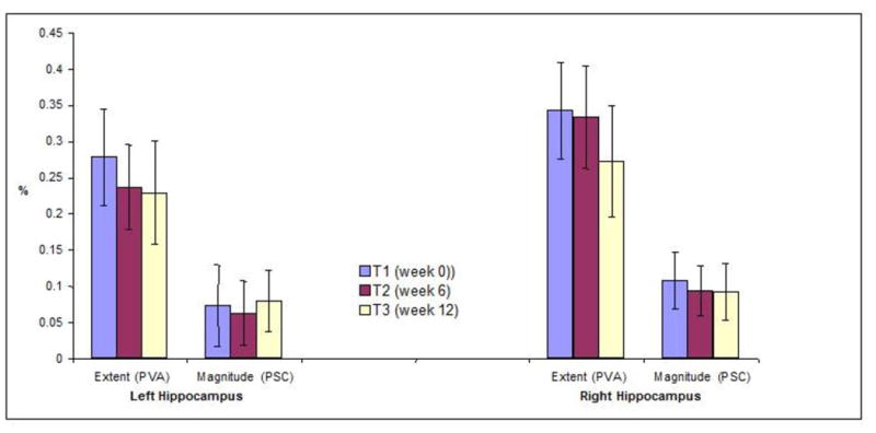
Main outcome measures: We performed whole-brain t maps (P < .001, 5 contiguous voxels) and hippocampal regions-of-interest analyses of extent (percentage of active voxels) and magnitude (percentage of signal change) for novel-greater-than-repeated face-name contrasts. We also calculated intraclass correlation coefficients and power estimates for hippocampal regions of interest.
Results: Task tolerability and data yield were high (95 of 96 scans yielded favorable-quality data). Whole-brain maps were stable. Right and left hippocampal regions-of-interest intraclass correlation coefficients were 0.59 to 0.87 and 0.67 to 0.74, respectively. To detect 25.0% to 50.0% changes in week-0 to week-12 hippocampal activity using left-right extent or right magnitude with 80.0% power (2-sided α = .05) requires 14 to 51 patients. Using left magnitude requires 125 patients because of relatively small signal to variance ratios.
Conclusions: Encoding-task fMRI was successfully implemented in a single-site, 24-week, AD randomized controlled trial. Week 0 to 12 whole-brain t maps were stable, and test-retest reliability of hippocampal fMRI measures ranged from moderate to substantial. Right hippocampal magnitude may be the most promising of these candidate measures in a leveraged context. These initial estimates of test-retest reliability and power justify evaluation of encoding-task fMRI as a potential biomarker for signal of effect in exploratory and proof-of-concept trials in mild AD. Validation of these results with larger sample sizes and assessment in multisite studies is warranted.
Figures
References
-
- Diamond EL, Miller S, Dickerson BC, et al. Relationship of fMRI activation to clinical trial memory measures in Alzheimer disease. Neurology. 2007 Sep 25;69(13):1331–1341. - PubMed
Publication types
MeSH terms
Substances
Grants and funding
LinkOut - more resources
Full Text Sources
Medical
