. 2011 Jun;121(6):2447-56.
doi: 10.1172/JCI46277. Epub 2011 May 9.

The histone trimethyllysine demethylase JMJD2A promotes cardiac hypertrophy in response to hypertrophic stimuli in mice

Affiliations

The histone trimethyllysine demethylase JMJD2A promotes cardiac hypertrophy in response to hypertrophic stimuli in mice

Qing-Jun Zhang et al. J Clin Invest. 2011 Jun.

Abstract

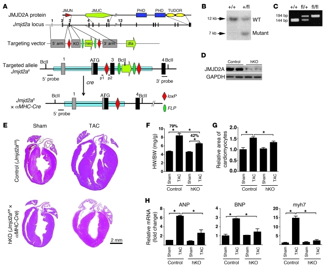
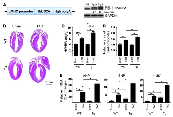
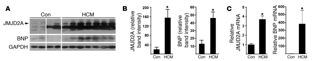
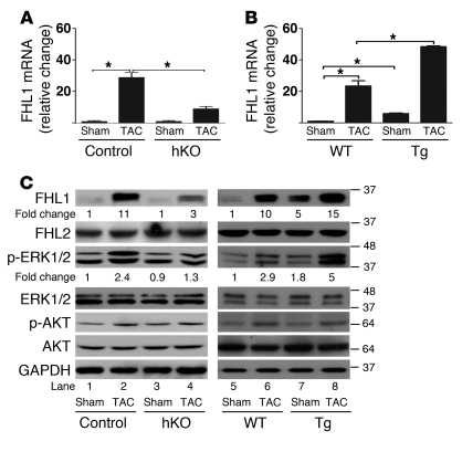
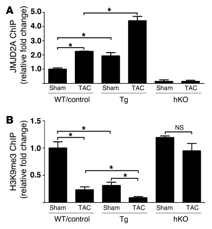
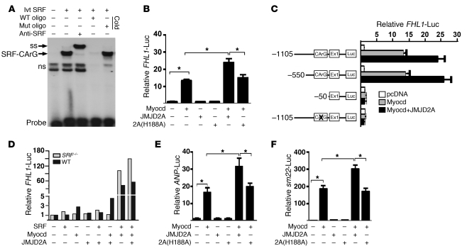
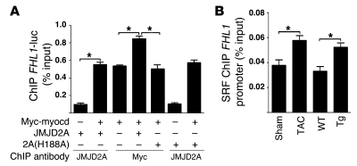
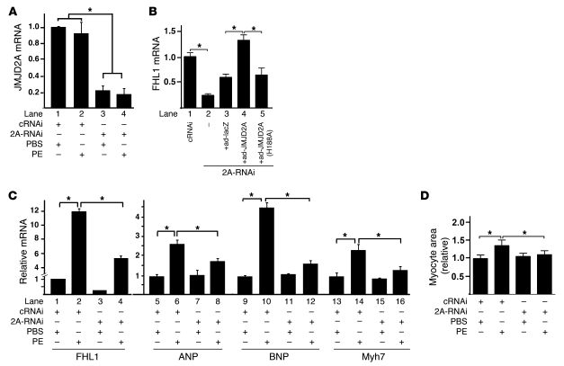
Cardiac hypertrophy and failure are accompanied by a reprogramming of gene expression that involves transcription factors and chromatin remodeling enzymes. Little is known about the roles of histone methylation and demethylation in this process. To understand the role of JMJD2A, a histone trimethyl demethylase, in cardiac hypertrophy, we generated mouse lines with heart-specific Jmjd2a deletion (hKO) and overexpression (Jmjd2a-Tg). Jmjd2a hKO and Jmjd2a-Tg mice had no overt baseline phenotype, but did demonstrate altered responses to cardiac stresses. While inactivation of Jmjd2a resulted in an attenuated hypertrophic response to transverse aortic constriction-induced (TAC-induced) pressure overload, Jmjd2a-Tg mice displayed exacerbated cardiac hypertrophy. We identified four-and-a-half LIM domains 1 (FHL1), a key component of the mechanotransducer machinery in the heart, as a direct target of JMJD2A. JMJD2A bound to the FHL1 promoter in response to TAC, upregulated FHL1 expression, and downregulated H3K9 trimethylation. Upregulation of FHL1 by JMJD2A was mediated through SRF and myocardin and required its demethylase activity. The expression of JMJD2A was upregulated in human hypertrophic cardiomyopathy patients. Our studies reveal that JMJD2A promotes cardiac hypertrophy under pathological conditions and suggest what we believe to be a novel mechanism for JMJD2A in reprogramming of gene expression involved in cardiac hypertrophy.


Figures

Figure 1
Figure 1. Generation and assessment of a cardiac-specific Jmjd2a KO mouse model.
(A) Strategy used to delete Jmjd2a in mouse cardiomyocytes and generate the Jmjd2a hKO model. Schematic protein structure of JMJD2A (14) and maps of the WT Jmjd2a locus, the targeting vector, the floxed allele, and the excised allele are shown. Exons are shown in bars. Exon 3 is flanked by loxP sites. (B) Southern blot of tail DNA from a WT (+/+) and a heterozygous (fl/+) mouse using 5′ probe shown in A after digestion with BclI. (C) PCR genotyping on genomic DNA isolated from a WT (+/+), a heterozygous (fl/+), and a homozygous floxed (fl/fl) mouse using primers p1/p2 indicated in A. (D) Western blot demonstrating significantly reduced JMJD2A protein in the heart homogenate of Jmjd2a hKO mice at 4 weeks. (E) H&E stains of representative paraffin sections of control and hKO hearts 3 weeks after sham and TAC operation. (F) HW/BW ratios of control and hKO mice 3 weeks after sham and TAC operation (n = 10–14 per group). The HW/BW ratio after TAC operation was increased 79% compared with sham operation in control mice, whereas it was only increased 42% in hKO mice. (G) Myocyte cross-sectional areas from control and hKO mice (>200 myocytes per heart in randomly selected filed) were assessed (n = 6–8 per group). (H) mRNA transcripts of ANP, BNP, and myh7 in control and hKO mouse hearts after 3 weeks sham and TAC operation. RNAs were normalized to internal GAPDH expression and presented as the fold change relative to control sham samples (n = 4–6 per group). Values are mean ± SEM. *P < 0.05.
Figure 2
Figure 2. JMJD2A promotes cardiac hypertrophy in response to TAC-induced pressure overload.
(A) Schematic diagram of cDNA encoding a flag-tagged JMJD2A in an expression vector containing the α-MHC promoter (left panel). 4 founder mice were obtained. 2 transgenic lines (Tg-A and Tg-B) with modest JMJD2A expression shown by Western blot analysis with anti-JMJD2A antibody (right panel) were established. The exogenous JMJD2A protein level is about 2-fold higher in the Tg-A line and 8-fold higher in the Tg-B line compared with that of endogenous JMJD2A. (B) H&E stains of paraffin section of WT and Jmjd2a-Tg (Tg) mouse hearts 3 weeks after sham and TAC operations. (C) HW/BW ratio (n = 6–7 per group) and (D) relative areas of cardiomyocytes (n = 3–4 per group) in WT and Jmjd2a-Tg sham- and TAC-operated animals. (E) Transcript levels of fetal gene markers, including ANP, BNP, and myh7, were quantified by real-time qRT-PCR, normalized against levels of internal GAPDH, and expressed as the fold change relative to that of sham-operated WT animals (n = 3 per group). The HW/BW ratio after TAC operation was increased 50% compared with sham operation in WT mice, whereas it was increased 100% in Tg mice. Values are mean ± SEM. *P < 0.05.
Figure 3
Figure 3. JMJD2A is upregulated in human hypertrophic hearts.
(A) Heart tissue samples of human HCM patients and unmatched non-HCM controls (con) were subjected to Western blot analysis using antibodies against JMJD2A, BNP, and GAPDH. Consistent with the HCM phenotype, BNP was upregulated in HCM samples. The lanes were run on the same gel but were noncontiguous. (B) More importantly, JMJD2A was significantly upregulated in HCM samples (n = 7) compared with samples from the non-HCM patients (n = 4). GAPDH was used as the loading control. (C) Relative mRNA levels of JMJD2A and BNP in control (n = 3) and HCM patients (n = 7) measured by qRT-PCR. Values are mean ± SEM. *P < 0.05.
Figure 4
Figure 4. JMJD2A upregulates the expression of FHL1.
qRT-PCR analysis of transcript levels of FHL1 from (A) control (Jmjd2afl/fl) and Jmjd2a hKO mouse hearts (n = 4–6) and (B) WT and Jmjd2a-Tg mouse hearts (n = 3–4) 21 days after sham and TAC surgery. Values are mean ± SEM. *P < 0.05. (C) Western blot of heart lysates of sham- and TAC-operated control and Jmjd2a hKO mice (left panel) and sham- and TAC-operated WT and Jmjd2a-Tg mice (right panel) using antibodies against FHL1, FHL2, phospho-ERK1/2, ERK1/2, phospho-AKT, and AKT. Representative of 4–6 experiments was shown. The relative fold changes for FHL1 and p-ERK were quantified by ImageJ. GAPDH was used as the loading control.
Figure 5
Figure 5. JMJD2A binds to the FHL1 promoter in vivo.
(A) JMJD2A-chromatin complexes were immunoprecipitated from lysates of sham- and TAC-operated WT, control (Jmjd2afl/fl), Jmjd2a-Tg, and Jmjd2a hKO mouse hearts, with anti-JMJD2A antibody and quantified by qPCR with primers in the FHL1 promoter. The amounts of immunoprecipitated complex were normalized against DNA purified from sonicated cell lysates (input) and expressed as relative to that of sham-operated WT/control. (B) ChIP was performed on aliquots of lysates from A using an anti-H3K9me3 antibody. The amounts of chromatin in the FHL1 promoter associated with H3K9me3 were measured by qPCR, normalized against input, and expressed relative to that of sham-operated WT/controls. JMJD2A binds the FHL1 promoter in response to TAC, and binding of JMJD2A is associated with decreased levels of H3K9me3. Values are expressed as mean ± SEM of 3 independent experiments. *P < 0.05.
Figure 6
Figure 6. JMJD2A upregulates FHL1 transcription synergistically with SRF/myocardin.
(A) A 32P-labeled oligonucleotide probe containing the CArG box from the FHL1 promoter was incubated with the in vitro–translated (ivt) SRF in the presence or absence of anti-SRF antibody or a 100-fold molar excess of unlabeled, WT, or mutant (mut) oligonucleotides. SRF forms a complex with the WT oligonucleotide probe (SRF-CArG) that can be super-shifted (ss) by an anti-SRF antibody. ns, nonspecific nucleoprotein complex. (B) A 1.9-kb FHL1 promoter–driven luciferase construct was transfected along with the plasmids indicated into QBI293A cells. FHL1-luciferase activities were measured 24 hours after transfection and normalized against cotransfected β-galactosidase. (C) Responsiveness of the deletion and site-specific mutants of the FHL1-luc reporter to myocardin and JMJD2A in QBI293A cells. Deletion or site-specific mutation of the SRF-binding site CArG box impairs the responsiveness of the promoter to myocardin and JMJD2A. (D) Relative activity of the FHL1-luc reporter in SRF-null and WT ES cells in the presence and absence of cotransfected expression plasmids as indicated. Representative of 3 independent experiments is shown. (E) Relative luciferase activity of the ANP-luc reporter and (F) the sm22-luc reporter in QBI293A cells transfected with the plasmids indicated. Myocardin-activated ANP- and sm22-luc reporters that can be further upregulated by WT but not mutant JMJD2A. Data shown are mean ± SEM of 3 independent experiments. *P < 0.05.
Figure 7
Figure 7. JMJD2A enhances the binding of SRF/myocardin to the FHL1 promoter.
(A) QBI293A cells were transfected with the plasmids indicated. ChIP assays were performed 24 hours later using anti-myc and anti-JMJD2A antibodies as indicated. (B) ChIP assays were performed with lysates of sham- and TAC-operated WT mouse hearts and with WT and Jmjd2a-Tg mouse hearts using anti-SRF antibody. Chromatins associated with SRF on the FHL1 promoter were quantified using qPCR. Data are expressed as mean ± SEM of 3 independent experiments. *P < 0.05.
Figure 8
Figure 8. Downregulation of JMJD2A attenuates the expression of FHL1 and PE-activated fetal gene program in cardiomyocytes.
Neonatal rat ventricular myocytes were transfected with nonspecific control siRNA (cRNAi) or JMJD2A-specific siRNA (2A-RNAi). Forty-eight hours after transfection, cells were treated with PBS (vehicle) or with PE (10 μM) (A, C, and D) or with various adenoviruses as indicated (B). Relative transcript levels of JMJD2A (A), FHL1 (B, lanes 1–5, and C, lanes 1–4), and fetal genes (C, lanes 5–16) were determined 24 hours after the treatment using qRT-PCR, normalized against internal GAPDH, and expressed relative to those of the vehicle-treated cRNAi transfected cells. (D) Cardiomyocytes were photographed, and areas were quantified using ImageJ and expressed relative to those of the vehicle-treated cRNAi transfected cells. Data are expressed as mean ± SEM of 3 independent experiments. *P < 0.05.

References

    1. Levy D, Garrison RJ, Savage DD, Kannel WB, Castelli WP. Prognostic implications of echocardiographically determined left ventricular mass in the Framingham Heart Study. N Engl J Med. 1990;322(22):1561–1566. - PubMed
    1. Hill JA, Olson EN. Cardiac plasticity. N Engl J Med. 2008;358(13):1370–1380. doi: 10.1056/NEJMra072139. - DOI - PubMed
    1. Abraham WT, et al. Coordinate changes in Myosin heavy chain isoform gene expression are selectively associated with alterations in dilated cardiomyopathy phenotype. Mol Med. 2002;8(11):750–760. - PMC - PubMed
    1. Blaxall BC, Tschannen-Moran BM, Milano CA, Koch WJ. Differential gene expression and genomic patient stratification following left ventricular assist device support. Am J Coll Cardiol. 2003. 41 7 1096 1106 . 10.1016/S0735-1097(03)00043-3 - DOI - PubMed
    1. Meyer K, Zhang L. Fetal programming of cardiac function and disease. Reprod Sci. 2007;14(3):209–216. doi: 10.1177/1933719107302324. - DOI - PubMed

Publication types

MeSH terms