. 2012 Feb;22(2):346-59.
doi: 10.1038/cr.2011.80. Epub 2011 May 10.

The p97 ATPase associates with EEA1 to regulate the size of early endosomes

Affiliations

The p97 ATPase associates with EEA1 to regulate the size of early endosomes

Harish N Ramanathan et al. Cell Res. 2012 Feb.

Abstract

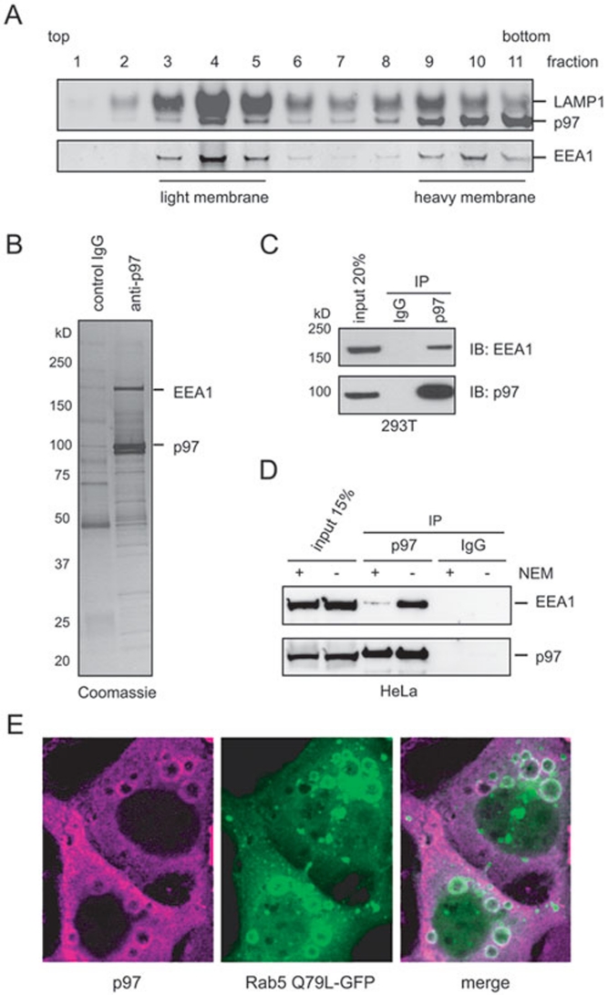
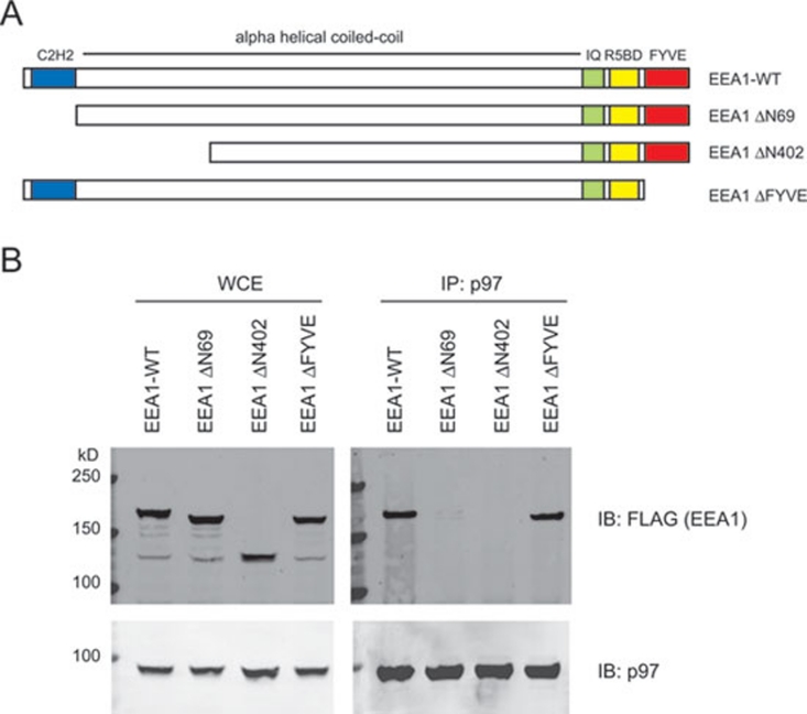
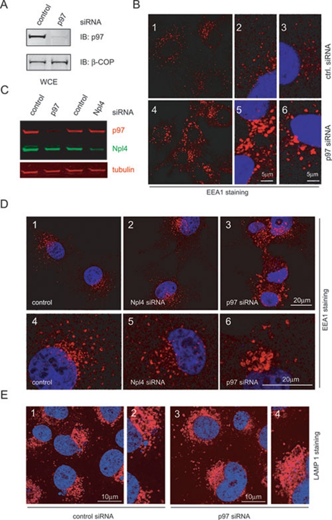
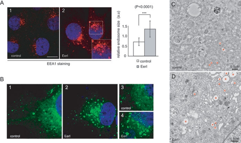
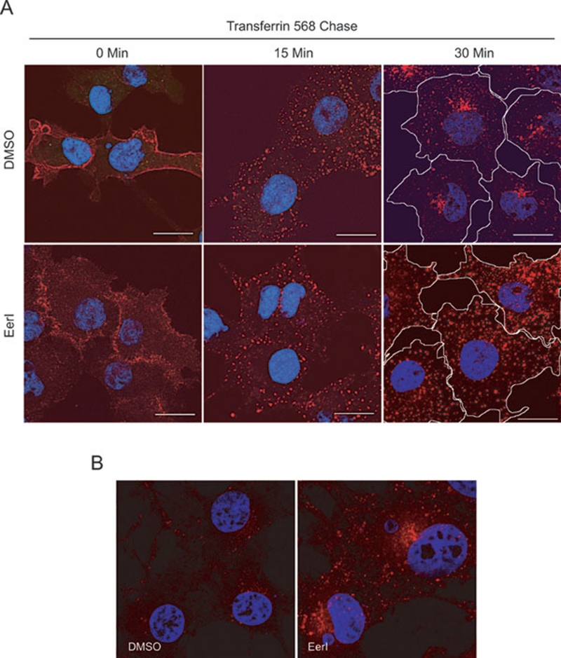
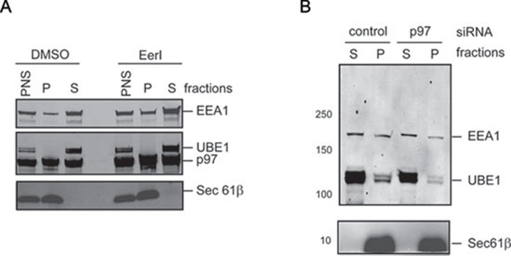
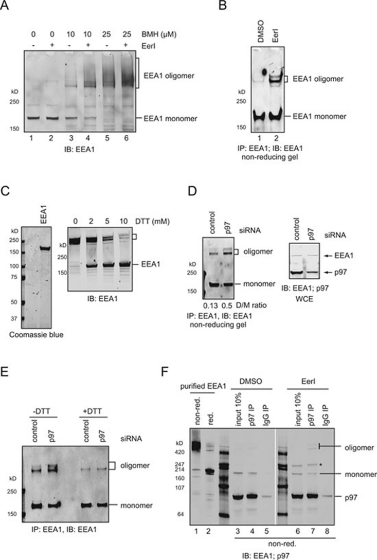
The AAA (ATPase-associated with various cellular activities) ATPase p97 acts on diverse substrate proteins to partake in various cellular processes such as membrane fusion and endoplasmic reticulum-associated degradation (ERAD). In membrane fusion, p97 is thought to function in analogy to the related ATPase NSF (N-ethylmaleimide-sensitive fusion protein), which promotes membrane fusion by disassembling a SNARE complex. In ERAD, p97 dislocates misfolded proteins from the ER membrane to facilitate their turnover by the proteasome. Here, we identify a novel function of p97 in endocytic trafficking by establishing the early endosomal autoantigen 1 (EEA1) as a new p97 substrate. We demonstrate that a fraction of p97 is localized to the early endosome membrane, where it binds EEA1 via the N-terminal C2H2 zinc finger domain. Inhibition of p97 either by siRNA or a pharmacological inhibitor results in clustering and enlargement of early endosomes, which is associated with an altered trafficking pattern for an endocytic cargo. Mechanistically, we show that p97 inhibition causes increased EEA1 self-association at the endosome membrane. We propose that p97 may regulate the size of early endosomes by governing the oligomeric state of EEA1.


Figures

Figure 1
Figure 1
The p97 is localized to the endosomes and interacts with EEA1. (A) A post nuclear supernatant fraction from 293T cells was subject to sucrose gradient floatation to separate membranes based on their density. Collected fractions were analyzed by immunoblotting with the indicated antibodies. (B) A Coomassie blue stained gel showing proteins co-purified with p97 from solubilized cow liver membrane. (C) Co-IP analysis of EEA1-p97 interaction using 293T cell extracts. (D) As in C, except that HeLa cells were used. Where indicated, cell extracts were treated with 5 mM NEM. (E) Confocal analyses of COS7 cells showing endogenous p97 (left panel), overexpressed Rab5 (Q79L) GFP (middle) and the merged image (right).
Figure 2
Figure 2
The C2H2 domain in EEA1 is required for p97 association. (A) Schematic representation of full-length EEA1 and the deletion constructs used in the binding study. IQ, calmodulin-binding site; R5BD, Rab5-binding domain. (B) Co-IP analysis of p97 interaction with the EEA1 variants. WCE, whole cell extract.
Figure 3
Figure 3
The p97 knockdown results in over-tethering/enlargement of early endosomes. (A) COS7 cells were treated with control or p97 specific siRNA. Whole cell extracts were subjected to immunoblotting to verify the knockdown efficiency. (B) Cells treated with control (panels 1, 2 and 3) or p97 specific siRNA (panels 4, 5 and 6) were immunostained to label endogenous EEA1 in red and DNA in blue. Panels 2, 3, 5 and 6 are close-up views of the cells. (C) COS7 cells were treated with control or p97-specific siRNA. Whole cell extracts were subjected to immunoblotting analysis with the indicated antibodies. (D) As in B, except that cells treated with the Npl4 siRNA were also included. Panels 4-6 are close-up views of the cells. (E) As in B, except that cells were stained with a LAMP1 antibody.
Figure 4
Figure 4
A p97 inhibitor causes enlargement of early endosomes. (A) COS7 cells treated with DMSO (control) or EerI (10 μM) for 6 h were stained for EEA1 in red and DNA in blue. The inset shows an enlarged view of the indicated region. Unless otherwise specified, scale bars correspond to 20 μm. The graph shows the quantification of the relative size of early endosomes in control and EerI-treated COS7 cells. a.u. arbitrary unit. Each value is the mean of 100 different vesicles, and the error bars represent the standard deviation. The p-value is obtained through paired t-test. (B) COS7 cells transfected with a low concentration of a Rab5-GFP-expressing plasmid were treated with either DMSO or EerI (10 μM) for 4 h. Cells were fixed and stained with DAPI. (C, D) COS7 cells treated with DMSO (C) or 10 μM EerI (D) for 4 h were fixed and analyzed by a transmission electron microscope at 4 000× magnification. Asterisks indicate examples of endosomes that are identified based on their unique membrane projections and lack of internal electron density .
Figure 5
Figure 5
Inhibition of p97 delays the trafficking of an endocytic cargo. (A) COS7 cells treated with DMSO or EerI (10 μM) for 1 h were labeled with Tfn and chased for 0, 15 and 30 min. Panels show the temporal distribution of Tfn in red relative to the nuclei in blue. In the 30 min panels, cell boundary is outlined in white. (B) As in A, except that cells chased for 60 min are shown. Note that in control cells, few Tfn-containing vesicles are detected due to recycling of Tfn.
Figure 6
Figure 6
The p97 does not regulate EEA1 membrane association. (A) COS7 cells treated with DMSO or EerI for 4 h were fractionated into membrane (P) and cytosolic (S) fractions. The relative distribution of the indicated proteins was analyzed by immunoblotting. (B) As in A, except that control and p97 knockdown cells were used.
Figure 7
Figure 7
Inhibition of p97 increases EEA1 self-association. (A) COS7 cells exposed to 10 μM EerI or DMSO for 4 h were treated by the indicated concentrations of BMH. WCE was analyzed by immunoblotting. (B) EEA1 was immunoprecipitated from detergent extracts of EerI- or DMSO-treated COS7 cells and analyzed by immunoblotting under a non-reducing condition. (C) Purified EEA1 was analyzed by SDS-PAGE under the reducing condition (left panel). Purified EEA1 that has been treated with the indicated concentration of the reducing agent DTT was analyzed by immunoblotting. (D) As in B, except that control and p97 knockdown cells were used. The bottom panel shows the knockdown of p97 in p97 siRNA treated cells. The numbers in the right panel indicate the ratio of EEA1 dimer to the monomer (D/M). (E) As in C. Where indicated, a fraction of the precipitated EEA1 was analyzed under the reducing condition (+DTT). (F) EEA1 co-immunoprecipitated with p97 from the indicated cells was analyzed by immunoblotting under the non-reducing condition (non-red.). Lanes 1 (non-reducing) and 2 (reducing) show purified EEA1 protein for comparison. The asterisk indicates a p97 oligomeric species present in EerI-treated cells .

Similar articles

Cited by

References

    1. DeLaBarre B, Brunger AT. Complete structure of p97/valosin-containing protein reveals communication between nucleotide domains. Nat Struct Biol. 2003;10:856–863. - PubMed
    1. Zhang X, Shaw A, Bates PA, et al. Structure of the AAA ATPase p97. Mol Cell. 2000;6:1473–1484. - PubMed
    1. Jentsch S, Rumpf S. Cdc48 (p97): a “molecular gearbox” in the ubiquitin pathway. Trends Biochem Sci. 2007;32:6–11. - PubMed
    1. Ye Y. Diverse functions with a common regulator: ubiquitin takes command of an AAA ATPase. J Struct Biol. 2006;156:29–40. - PubMed
    1. Wilcox AJ, Laney JD. A ubiquitin-selective AAA-ATPase mediates transcriptional switching by remodelling a repressor-promoter DNA complex. Nat Cell Biol. 2009;11:1481–1486. - PMC - PubMed

Publication types

MeSH terms