. 2011 May 10;30(12):2325-35.
doi: 10.1038/emboj.2011.157.

Modulating F-actin organization induces organ growth by affecting the Hippo pathway

Affiliations

Modulating F-actin organization induces organ growth by affecting the Hippo pathway

Leticia Sansores-Garcia et al. EMBO J. .

Abstract

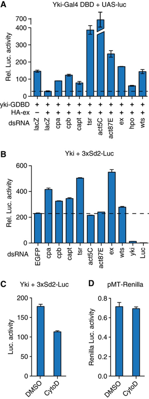
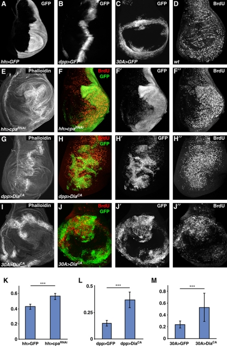
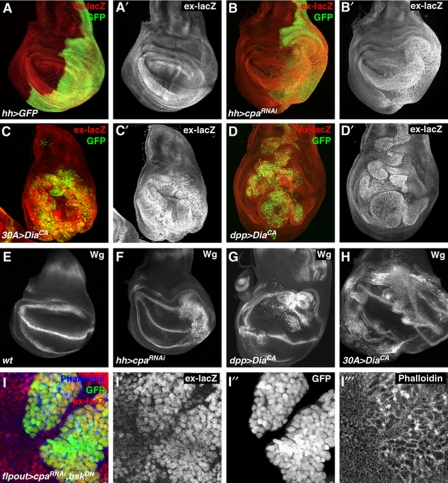
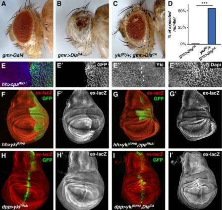
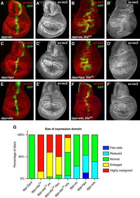
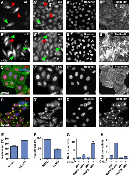
The Hippo tumour suppressor pathway is a conserved signalling pathway that controls organ size. The core of the Hpo pathway is a kinase cascade, which in Drosophila involves the Hpo and Warts kinases that negatively regulate the activity of the transcriptional coactivator Yorkie. Although several additional components of the Hippo pathway have been discovered, the inputs that regulate Hippo signalling are not fully understood. Here, we report that induction of extra F-actin formation, by loss of Capping proteins A or B, or caused by overexpression of an activated version of the formin Diaphanous, induced strong overgrowth in Drosophila imaginal discs through modulating the activity of the Hippo pathway. Importantly, loss of Capping proteins and Diaphanous overexpression did not significantly affect cell polarity and other signalling pathways, including Hedgehog and Decapentaplegic signalling. The interaction between F-actin and Hpo signalling is evolutionarily conserved, as the activity of the mammalian Yorkie-orthologue Yap is modulated by changes in F-actin. Thus, regulators of F-actin, and in particular Capping proteins, are essential for proper growth control by affecting Hippo signalling.


Conflict of interest statement

The authors declare that they have no conflict of interest.

Figures

Figure 1
Figure 1
Actin modulators regulate Yorkie activity in vitro. Luciferase activity assays normalized with a Renilla luciferase control for transfection efficiency. (A) Drosophila S2 cells were transiently transfected with plasmids expressing UAS-Luciferase and Yki-GDBD alone or cotransfected with HA-Ex constructs in the presence of the dsRNAs as indicated. (B) Drosophila S2 cells were transiently transfected with the Yki-reporter construct 3xSd2-Luciferase and pMT-Yki in the presence of the dsRNAs as indicated. (C) Luciferase activity produced by pMT-Yki driving the 3xSd2-Luciferase reporter construct in vehicle (DMSO) and cytochalasin D (CytoD) treated S2 cells. (D) Renilla Luciferase activity produced by the transfection control plasmid is the same in vehicle (DMSO) and cytochalasin D (CytoD) treated S2 cells. The error bars represent ‘standard error of the mean’ (s.e.m.).
Figure 2
Figure 2
Induction of F-actin polymerization induces overgrowth of imaginal discs. (AJ) Confocal images of third instar wing imaginal discs showing (A) UAS-GFP expression driven by hh-Gal4, (B) UAS-GFP expression driven by dpp-Gal4, and (C) UAS-GFP expression driven by 30A-Gal4. (D) BrdU incorporation in a wild-type disc. (E) Phalloidin staining of hh-Gal4, UAS-GFP, UAS-cpaRNAi. (F) BrdU (red, grey in F′′) and GFP (green, grey in F′) staining of hh-Gal4, UAS-GFP, UAS-cpaRNAi. (G) Phalloidin staining of dpp-Gal4, UAS-GFP, UAS-diaCA. (H) BrdU (red, grey in H′′) and GFP (green, grey in H′) staining of dpp-Gal4, UAS-GFP, UAS-diaCA. (I) Phalloidin staining of 30A-Gal4, UAS-GFP, UAS-diaCA. (J) BrdU (red, grey in J′′) and GFP (green, grey in J′) staining of 30A-Gal4, UAS-GFP, UAS-diaCA. (KM) Quantification of the size of the GFP expression domains normalized to the size of the entire imaginal disc of the indicated genotypes. *** indicates that the two populations are different with P<0.001. Note that Gal4 in the dpp- and 30A-Gal4 driver lines is not stably expressed in cell lineages but is lost when progenitor cells move away from the domain where Gal4 expression was induced. Therefore, non-GFP expressing cells may show upregulated BrdU due to earlier Gal4 expression driving DiaCA. Thus, seemingly non-autonomous effects may still be due to cell autonomous action of DiaCA. Similar effects may apply to analysis of the expression of ex-lacZ in Figure 3.
Figure 3
Figure 3
Actin modulators regulate Hippo pathway target genes. Confocal images of third instar wing imaginal discs. (AD) GFP (green) and ex-lacZ (red, grey in A′D′) staining of (A) hh-Gal4, UAS-GFP, (B) hh-Gal4, UAS-GFP, UAS-cpaRNAi, (C) 30A-Gal4, UAS-GFP, UAS-diaCA, and (D) dpp-Gal4, UAS-GFP, UAS-diaCA. (EH) Wingless (Wg) antibody staining of (E) wild type, (F) hh-Gal4, UAS-GFP, UAS-cpaRNAi, (G) dpp-Gal4, UAS-GFP, UAS-diaCA, and (H) 30A-Gal4, UAS-GFP, UAS-diaCA. (I) Close-up image of a clone of cells in the wing pouch region of a wing disc expressing UAS-cpaRNAi, UAS-bskDN, and UAS-GFP driven by Flip-out-Gal4 stained for ex-lacZ (red, grey in I′) and Phalloidin (blue, grey in I′′′) and showing GFP expression (green, grey in I′′).
Figure 4
Figure 4
Yorkie is required for actin dynamics induced overgrowth and upregulation of Hippo target genes. (AC) Adult flies of the indicated genotypes. (D) Quantification of the number of enclosed adult flies of the genotypes in (B, C) relative to (A). (E) Confocal image of a third instar wing imaginal disc stained for GFP (green, grey in E′), Yki (red, grey in E′′), and Dapi (blue, grey in E′′′) of hh-Gal4, UAS-GFP, UAS-cpaRNAi. (F, I) Third instar wing imaginal discs stained for GFP (green) and ex-lacZ (red, grey in F′I′) of (F) hh-Gal4, UAS-GFP, UAS-ykiRNAi, (G) hh-Gal4, UAS-GFP, UAS-ykiRNAi, UAS-cpaRNAi, (H) dpp-Gal4, UAS-GFP, UAS-ykiRNAi, and (I) dpp-Gal4, UAS-GFP, UAS-ykiRNAi, UAS-diaCA.
Figure 5
Figure 5
Overexpression of Warts suppresses Diaphanous-induced phenotypes. Confocal images of ex-lacZ (red and grey A′F′) and GFP (green) stainings of third instar wing imaginal discs of (A) dpp-Gal4, UAS-GFP, UAS-ex, (B) dpp-Gal4, UAS-GFP, UAS-ex, UAS-diaCA, (C) dpp-Gal4, UAS-GFP, UAS-hpo, (D) dpp-Gal4, UAS-GFP, UAS-hpo, UAS-diaCA, (E) dpp-Gal4, UAS-GFP, UAS-wts, and (F) dpp-Gal4, UAS-GFP, UAS-wts, UAS-diaCA. (G) Semi-quantification of the sizes of the expression domains of discs with the genotypes shown in this figure.
Figure 6
Figure 6
Actin dynamics regulate Hippo signalling in mammalian HeLa cells. (AD) In vitro culture of HeLa cells. (A) GFP construct transfection as a control and staining of (A) GFP, (A′) Yap, (A′′) Hoechst, and (A′′′) Phalloidin. Red arrowheads indicate dispersed Yap staining, green arrowheads indicate nuclear Yap staining. (B) Cotransfection of GFP and mDiaCA and staining for (B) GFP, (B′) Yap, (B′′) Hoechst, and (B′′′) Phalloidin. (C) Cells treated with vehicle (DMSO) and stained for (C′) Yap, (C′′) Hoechst, and (C′′′) Phalloidin. (D) Cells treated with cytochalasin D (CytoD) and stained for (D′) Yap, (D′′) Hoechst, and (D′′′) Phalloidin. (E) Quantification of nuclear localization of Yap upon transfection with a plasmid expressing mDiaCA. (F) Quantification of nuclear localization of Yap upon CytoD treatment. (G) Quantification of Luciferase expression from a TEAD-response luciferase construct (8xGTIIc) in the presence and absence of mDiaCA expression. (H) Quantification of Luciferase expression from a TEAD-response luciferase construct (8xGTIIc) with vehicle or CytoD treatment. The error bars in all graphs represent ‘standard error of the mean’ (s.e.m.).

Similar articles

Cited by

References

    1. Assoian RK, Klein EA (2008) Growth control by intracellular tension and extracellular stiffness. Trends Cell Biol 18: 347–352 - PMC - PubMed
    1. Bazellieres E, Assemat E, Arsanto JP, Le Bivic A, Massey-Harroche D (2009) Crumbs proteins in epithelial morphogenesis. Front Biosci 14: 2149–2169 - PubMed
    1. Benlali A, Draskovic I, Hazelett DJ, Treisman JE (2000) Act up controls actin polymerization to alter cell shape and restrict Hedgehog signaling in the Drosophila eye disc. Cell 101: 271–281 - PubMed
    1. Bennett FC, Harvey KF (2006) Fat cadherin modulates organ size in Drosophila via the Salvador/Warts/Hippo signaling pathway. Curr Biol 16: 2101–2110 - PubMed
    1. Boedigheimer M, Laughon A (1993) Expanded: a gene involved in the control of cell proliferation in imaginal discs. Development 118: 1291–1301 - PubMed

MeSH terms

LinkOut - more resources