. 2011 May 10;30(12):2405-19.
doi: 10.1038/emboj.2011.154.

Phospho-MED1-enhanced UBE2C locus looping drives castration-resistant prostate cancer growth

Affiliations

Phospho-MED1-enhanced UBE2C locus looping drives castration-resistant prostate cancer growth

Zhong Chen et al. EMBO J. .

Abstract

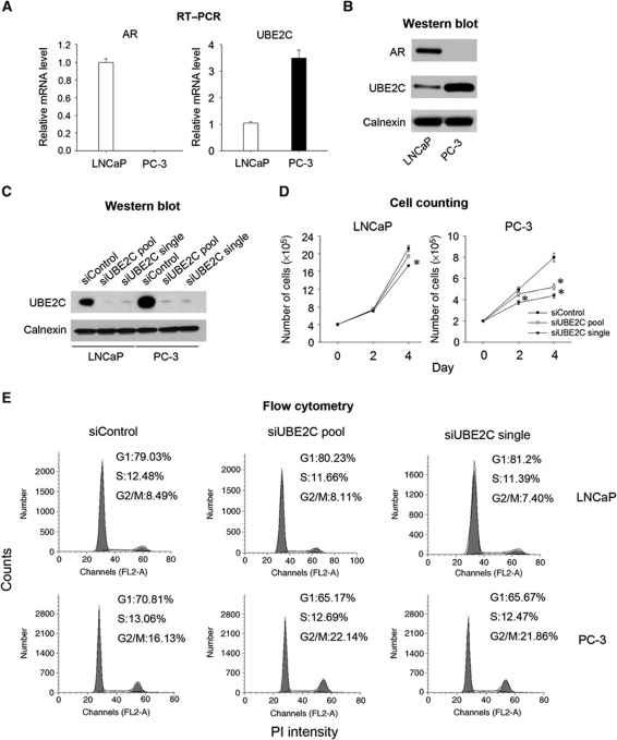
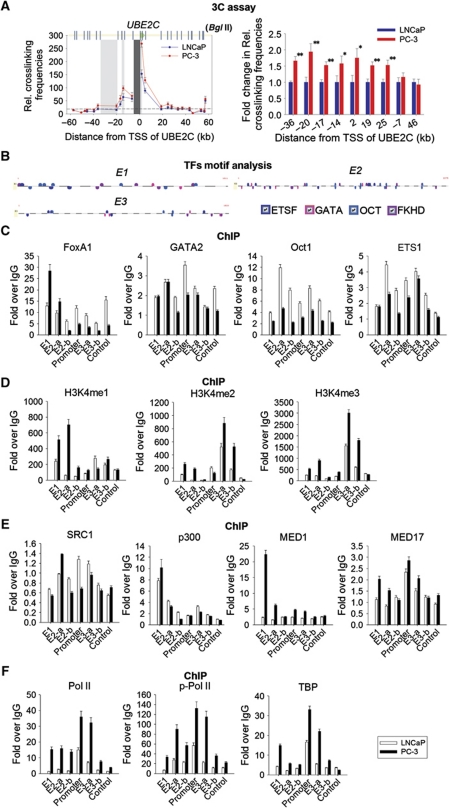
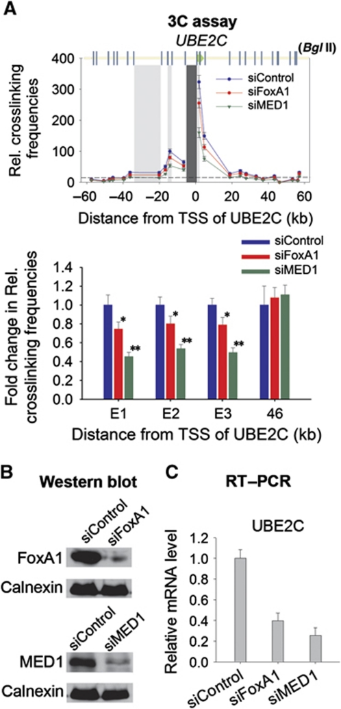
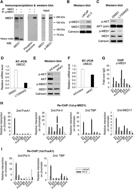
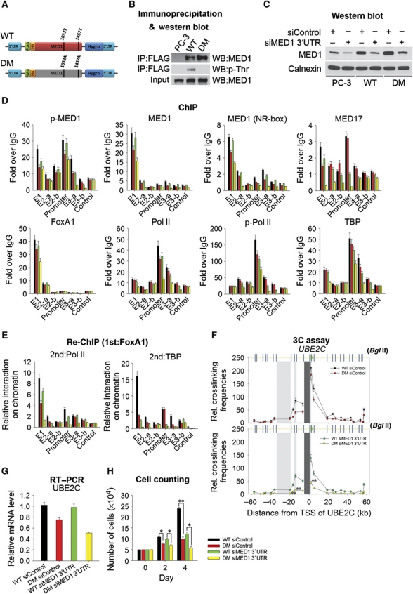
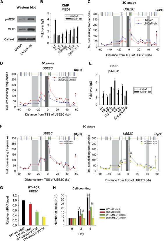
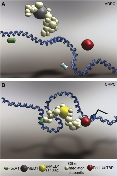
The UBE2C oncogene is overexpressed in many types of solid tumours including the lethal castration-resistant prostate cancer (CRPC). The underlying mechanisms causing UBE2C gene overexpression in CRPC are not fully understood. Here, we show that CRPC-specific enhancers drive UBE2C overexpression in both AR-negative and -positive CRPC cells. We further show that co-activator MED1 recruitment to the UBE2C enhancers is required for long-range UBE2C enhancer/promoter interactions. Importantly, we find that the molecular mechanism underlying MED1-mediated chromatin looping involves PI3K/AKT phosphorylated MED1-mediated recruitment of FoxA1, RNA polymerase II and TATA binding protein and their subsequent interactions at the UBE2C locus. MED1 phosphorylation leads to UBE2C locus looping, UBE2C gene expression and cell growth. Our results not only define a causal role of a post-translational modification (phosphorylation) of a co-activator (MED1) in forming or sustaining an active chromatin structure, but also suggest that development of specific therapies for CRPC should take account of targeting phosphorylated MED1.


Conflict of interest statement

The authors declare that they have no conflict of interest.

Figures

Figure 1
Figure 1
Increased UBE2C expression is required for PC-3 cell growth. (A) The UBE2C mRNA level is higher in PC-3 cells than in LNCaP cells. Total RNA was isolated and amplified with primers recognizing AR mRNA and UBE2C mRNA (mean (n=3)±s.d.). (B) The UBE2C protein expression level is higher in PC-3 cells than in LNCaP cells. Western blots analyses were performed using the indicated antibodies. (C) Suppression of UBE2C protein levels by RNAi. LNCaP and PC-3 cells were transfected with a siUBE2C ON-TARGET pool including four siRNAs and a single siUBE2C not included in the siUBE2C ON-TARGET pool, and western blots were performed using an anti-UBE2C antibody. (D) UBE2C silencing significantly decreases PC-3 growth. The cell numbers were determined by a direct viable cell count assay (mean (n=2)±s.d.) (two-sided t-test, *P<0.05). (E) UBE2C silencing arrests cell cycle in G2/M-phase. Ninety-six hours after UBE2C siRNAs transfection, LNCaP and PC-3 cells were analysed by FACS. A representative result of three independent experiments was shown.
Figure 2
Figure 2
Identification and characterization of UBE2C enhancers in PC-3 cells. (A) Greater interactions between seven distal regions and the UBE2C promoter in PC-3 cells than in LNCaP cells. Left panel: 3C assays were performed in LNCaP and PC-3 cells in the absence of DHT. The black shading shows the position of the fixed fragment (the UBE2C promoter). The grey shading indicates three fragments that were selected for further functional analysis. Right panel: The results of 3C assays were presented as fold changes in relative crosslinking frequencies (two-sided t-test, *P<0.05, **P<0.01). The −7 and +46 kb regions were used as controls. (B) Schematic representation of ETSF, GATA, OCT and FKHD motifs within the E1, E2 and E3 regions. (C) Increased FoxA1 binding at the UBE2C enhancers in PC-3 cells than in LNCaP cells. ChIP assays were performed using antibodies against FoxA1, GATA2, Oct1 and ETS1 in LNCaP and PC-3 cells in the absence DHT (mean (n=3)±s.d.). (D) Higher levels of H3K4 methylation on the UBE2C enhancers in PC-3 cells compared with LNCaP cells. ChIP assays were performed as above using antibodies against H3K4me1, H3K4me2 and H3K4 me3 (mean (n=3)±s.d.). (E) Increased recruitment of MED1 and/or MED17 to the UBE2C enhancers and promoter. ChIP assays were conducted as above using SRC1, p300, MED1 and MED17 antibodies. (F) Higher levels of Pol II and TBP occupancy on the UBE2C promoter and enhancers in PC-3 cells than in LNCaP cells. ChIP assays were performed as above using antibodies against Pol II, phosphorylated Pol II and TBP (mean (n=3)±s.d.).
Figure 3
Figure 3
Silencing of FoxA1 and MED1 impairs long-range interactions at the UBE2C locus and decreases UBE2C gene expression in PC-3 cells. (A) Upper panel: knocking down of FoxA1 and MED1 decreases crosslinking frequencies between the UBE2C enhancers and the UBE2C promoter. 3C assays were performed in siControl, siFoxA1 or siMED1 transfected PC-3 cells. Lower panel: the 3C results were presented as fold changes in relative crosslinking frequencies (two-sided t-test, *P<0.05, **P<0.01). (B) Suppression of FoxA1 and MED1 levels by siRNAs. PC-3 cells were transiently transfected with siControl, siFoxA1 or siMED1 and protein levels were determined by western blot analysis. (C) Silencing of FoxA1 and MED1 decreases UBE2C gene expression in PC-3 cells. PC-3 cells were transfected with siRNA targeting FoxA1 and MED1. Seventy-two hours after siRNA transfection, real-time RT–PCR was performed (mean (n=3)±s.d.).
Figure 4
Figure 4
Enhanced interactions between PI3K/AKT-induced T1032 phosphorylated MED1 and UBE2C enhancer/promoter-bound proteins on chromatin in PC-3 cells. (A) Characterization of a phosphorylated T1032-specific MED1 (p-MED1) antibody. Whole cell lysates from PC-3 cells were immunoprecipitated with a commercial MED1 antibody or p-MED1. Western blot analyses were then performed using indicated antibodies. (B) Comparisons of phosphorylated MED1 and MED1 protein levels in LNCaP and PC-3 cells. Western blot analyses were performed with the p-MED1 antibody. The same membrane was then reprobed with the MED1 antibody. (C) Effects of LY294002 on MED1 phosphorylation at T1032. PC-3 cells were treated with 50 μM LY294002 or vehicle for 24 h, and western blot analyses were performed using antibodies indicated. (D) Effects of LY294002 on UBE2C mRNA expression. Total RNA was isolated from LY294002 or vehicle-treated PC-3 cells, and amplified with UBE2C primers (mean (n=3)±s.d.). (E) Silencing of AKT decreases MED1 phosphorylation at T1032 in PC-3 cells. Seventy-two hours after siRNA transfection, western blot analyses were performed using antibodies indicated. (F) AKT silencing decreases UBE2C mRNA expression. Real-time RT–PCR was performed 72 h after sRNA transfection (mean (n=3)±s.d.). (G) Higher recruitment of phosphorylated MED1 to the UBE2C enhancers and promoter in PC-3 cells than in LNCaP cells. ChIP assays were conducted with the p-MED1 antibody (mean (n=3)±s.d.). (H) Increased protein–protein interactions between phosphorylated MED1 with UBE2C enhancer and promoter-bound proteins in PC-3 cells compared with LNCaP cells. ChIPs were first performed with the p-MED1 antibody. The immunoprecipitated complexes were eluted and the second ChIPs were performed with antibodies against FoxA1, Pol II, TBP and MED17 (mean (n=3)±s.d.). (I) Increased protein–protein interactions between FoxA1 and Pol II/TBP. Re-ChIP assays were performed using antibodies against FoxA1 (for first ChIP) and Pol II/TBP (for second ChIP).
Figure 5
Figure 5
Phosphorylation of MED1 in PC-3 cells is required for UBE2C locus looping, UBE2C gene expression and cell growth. (A) Schematic diagram of the retroviral vectors expressing FLAG/HA-tagged wild-type (WT) MED1 and T1032A/T1457A double-mutated (DM) MED1. (B) Immunoprecipitation of epitope-tagged WT MED1 and DM MED1 from PC-3/WT MED1 and PC-3/DM MED1 cells. Whole cell lysates were immunoprecipitated with an anti-FLAG antibody. Western blot analyses were then performed using an anti-MED1 antibody and an anti-phospho-threonine antibody. (C) Suppression of MED1 expression by RNAi. Parental PC-3, PC-3/WT MED1 and PC-3/DM MED1 cells were transfected with siMED1 targeting MED1 3′UTR, and western blot was performed with an anti-MED1 antibody. (D) Decreased recruitment of proteins to the UBE2C locus in PC-3/DM MED1 cells compared with PC-3/WT MED1 cells. PC-3/DM MED1 cells and PC-3/WT MED1 cells were transfected with siControl and siMED1 3′UTR. ChIP assays were performed using antibodies indicated (mean (n=3)±s.d.). (E) Decreased interactions between FoxA1 and Pol II/TBP at the UBE2C locus in PC-3/DM MED1 cells compared with PC-3/WT MED1 cells. PC-3/DM MED1 cells and PC-3/WT MED1 cells were transfected with siControl and siMED1 3′UTR. Re-ChIP assays were conducted with antibodies against FoxA1 (for first ChIP) and Pol II/TBP (for re-ChIP) (mean (n=3)±s.d.). (F) Disrupted UBE2C locus looping in PC-3/DM MED1 cells compared with PC-3/WT MED1 cells (two-sided t-test, **P<0.01). 3C assays were performed using siControl and siMED1 3′UTR transfected PC-3/DM MED1 cells and PC-3/WT MED1 cells. (G) UBE2C mRNA expression is lower in PC-3/DM MED1 cells than in PC-3/WT MED1 cells transfected with siMED1 3′UTR (mean (n=3)±s.d.). (H) PC-3/DM MED1 cells transfected with siMED1 3′UTR grow slower than PC-3/WT MED1 cells transfected with siMED1 3′UTR. The cell proliferation was measured using a direct viable cell count assay (mean (n=2)±s.d.) (two-sided t-test, *P<0.05, **P<0.01).
Figure 6
Figure 6
Phosphorylated MED1 in LNCaP-abl cells enhances UBE2C locus looping, UBE2C mRNA expression and cell growth. (A) Comparison of MED1 and phosphorylated MED1 expression levels in LNCaP and LNCaP-abl cells. Western blot analyses were performed using the antibodies indicated. (B) Increased MED1 binding at the UBE2C enhancers and UBE2C promoter in LNCaP-abl compared with LNCaP cells. ChIP assays were performed using an anti-MED1 antibody (mean (n=3)±s.d.). (C) Increased interactions between the UBE2C enhancers (E1, E2, E3 and Enhancer-2) and the UBE2C promoter in LNCaP-abl compared with LNCaP cells. 3C assays were performed in LNCaP-abl and LNCaP cells in the absence of DHT. (D) Silencing of MED1 decreases crosslinking frequencies between the UBE2C enhancers and the UBE2C promoter in LNCaP-abl cells. 3C assays were performed in siControl or siMED1 transfected LNCaP-abl cells. (E) Increased recruitment of phosphorylated MED1 to the UBE2C regulatory regions in LNCaP-abl compared with LNCaP cells. ChIP assays were performed with the p-MED1 antibody. (F) Disrupted UBE2C locus looping in LNCaP-abl/DM MED1 cells compared with LNCaP/WT MED1 cells (two-sided t-test *P<0.05, **P<0.01). 3C assays were performed following siControl or siMED1 3′UTR transfection. (G) Decreased UBE2C mRNA expression in LNCaP-abl/DM MED1 cells transfected with siMED1 3′UTR compared with LNCaP-abl/WT MED1 cells transfected with siMED1 3′UTR (mean (n=3)±s.d.). (H) Decreased cell growth of LNCaP-abl/DM MED1 cells transfected with siMED1 3′UTR compared with LNCaP-abl/WT MED1 cells transfected with siMED1 3′UTR. (mean (n=2)±s.d.) (two-sided t-test, *P<0.05, **P<0.01).
Figure 7
Figure 7
Models of phosphorylated MED1-mediated chromatin looping. (A) In ADPC cells, unphosphorylated MED1 does not mediate recruitment of FoxA1, Pol II and TBP and their subsequent interactions on chromatin. (B) In CRPC cells, phosphorylated MED1 at T1032 facilitates FoxA1, Pol II and TBP recruitment and mediates their interactions on chromatin leading to chromatin looping.

Similar articles

Cited by

References

    1. Barski A, Cuddapah S, Cui K, Roh TY, Schones DE, Wang Z, Wei G, Chepelev I, Zhao K (2007) High-resolution profiling of histone methylations in the human genome. Cell 129: 823–837 - PubMed
    1. Basu S, Totty NF, Irwin MS, Sudol M, Downward J (2003) Akt phosphorylates the Yes-associated protein, YAP, to induce interaction with 14-3-3 and attenuation of p73-mediated apoptosis. Mol Cell 11: 11–23 - PubMed
    1. Belakavadi M, Pandey PK, Vijayvargia R, Fondell JD (2008) MED1 phosphorylation promotes its association with mediator: implications for nuclear receptor signaling. Mol Cell Biol 28: 3932–3942 - PMC - PubMed
    1. Bolton EC, So AY, Chaivorapol C, Haqq CM, Li H, Yamamoto KR (2007) Cell- and gene-specific regulation of primary target genes by the androgen receptor. Genes Dev 21: 2005–2017 - PMC - PubMed
    1. Bulger M, Groudine M (2011) Functional and mechanistic diversity of distal transcription enhancers. Cell 144: 327–339 - PMC - PubMed

Publication types

MeSH terms