. 2011:2:308.
doi: 10.1038/ncomms1306.

Chemical treatment enhances skipping of a mutated exon in the dystrophin gene

Affiliations
Free PMC article

Chemical treatment enhances skipping of a mutated exon in the dystrophin gene

Atsushi Nishida et al. Nat Commun. 2011.
Free PMC article

Abstract

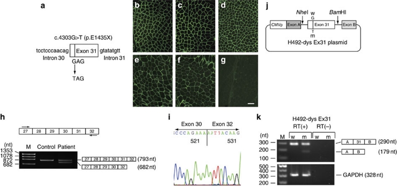
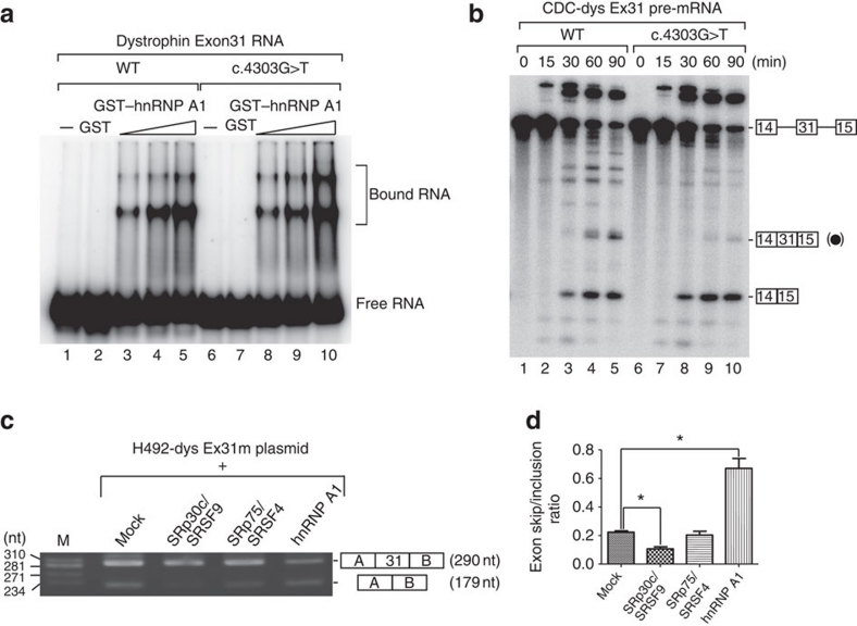
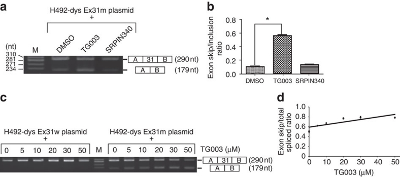
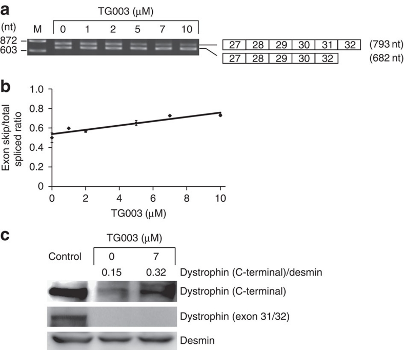
Duchenne muscular dystrophy (DMD) is a fatal muscle wasting disease caused by a loss of the dystrophin protein. Control of dystrophin mRNA splicing to convert severe DMD to a milder phenotype is attracting much attention. Here we report a dystrophinopathy patient who has a point mutation in exon 31 of the dystrophin gene. Although the mutation generates a stop codon, a small amount of internally deleted, but functional, dystrophin protein is produced in the patient cells. An analysis of the mRNA reveals that the mutation promotes exon skipping and restores the open reading frame of dystrophin. Presumably, the mutation disrupts an exonic splicing enhancer and creates an exonic splicing silencer. Therefore, we searched for small chemicals that enhance exon skipping, and found that TG003 promotes the skipping of exon 31 in the endogenous dystrophin gene in a dose-dependent manner and increases the production of the dystrophin protein in the patient's cells.


Figures

Figure 1
Figure 1. Point mutation in exon 31 of the dystrophin gene causes exon skipping and restores the open reading frame for an internally deleted protein.
(a) A point mutation that was found in the dystrophin gene of patient KUCG797. The position of c.4303G>T (p.Glu1435X) in exon 31 is indicated by the bar. (bg) Dystrophin expression in the control (bd) and in the patient's (eg) muscles. Immunohistochemical examinations for C-terminal (DYS2), N-terminal (DYS3) and rod- (MANDYS1) domains were carried out. Immunoreactivity for DYS2 (exons 77–79 of dystrophin) and DYS3 (exons 10–12 of dystrophin) (e, f) was somewhat patchy and weaker in the patient's muscles as compared with the control cells (b, c). However, immunoreactivity for MANDYS1, which recognizes exon 31/32 of the dystrophin was defective only in the patient (compare panels d, g). Scale bar, 50 μm. (h) The RT–PCR products obtained from the control and patient were analysed on agarose gels. The RT–PCR products from the patient's RNA contained an additional shorter product. The DNA sequences of these bands were analysed, and the structure of each PCR product is shown schematically at the right of the panel. (i) Sequencing of the shorter product seen in (c) confirmed the skipping of exon 31 in the patient. (j) A schematic representation of the hybrid minigene plasmids that harbour either the wild-type (W) or mutated (m) exon 31 of the dystrophin gene. The minigene vector H492 encodes two cassette exons (A and B) and an intron sequence containing a multicloning site. The dystrophin gene region encompassing exon 31 with flanking introns was inserted into the NheI and BamHI sites in the intron region of H492. These plasmids were transfected into HeLa cells and the pre-mRNAs were transcribed from the cytomegalovirus (CMV) promoter (CMVp). (k) The RT–PCR products of wild-type (W) and mutant (m) mRNA that were recovered from transfected HeLa cells were visualized on agarose gel. Two different PCR products were detected only with H492-dys Ex31m plasmid. Glyceraldehyde-3-phosphate dehydrogenase (GAPDH) mRNA was used as a control for transfection efficiency. As negative controls, RT–PCR reactions without reverse transcriptase (RT−) were performed for both RNAs from the reporters and GAPDH.
Figure 2
Figure 2. The point mutation in exon 31 enhances binding to hnRNP A1 and exon skipping both in vitro and in vivo.
(a) Gel mobility shift assays with GST–hnRNP A1 and dystrophin exon 31 RNA. 32P-labelled dystrophin exon 31 RNA (wild type or mutant) was incubated with either GST alone (GST: lanes 2 and 7, 400 ng) or with GST-tagged hnRNP A1 (GST–hnRNP A1: lanes 3–5 and 8–10; 100, 200 and 400 ng, respectively), and the resultant complexes were subjected to 8% native polyacrylamide gel electrophoresis. Lanes 1 and 6 show where RNA itself migrates on the gel (marked as Free RNA on the right of the panel). Complexes of hnRNP A1 and RNA are also indicated as Bound RNA. All incubations were carried out at 20 °C for 30 min. (b) In vitro splicing assays with pre-mRNAs that contain exon 31. 32P-labelled chicken δ-crystallin (CDC) pre-mRNA containing either wild-type (WT) or mutant (c.4303G>T) exon 31 in the intron was incubated with HeLa cell nuclear extracts at 30 °C for the time shown at the top of the panel. The RNA products were analysed by electrophoresis on 6% denaturing gel. The structures of the pre-mRNA and two different mRNAs are shown at the right of the panel. Boxes with numbers indicate exons, whereas the lines between boxes designate introns. The mRNA that includes exon 31 (closed circle) was produced more efficiently with CDC-dys Ex31 WT pre-mRNA than CDC-dys Ex31 c.4303G>T pre-mRNA. (c) Effect of overexpression of several RNA-binding proteins on the inclusion and skipping of the mutant exon 31. HeLa cells (3×10⁶ cells) were cultured overnight and transfected with 150 ng of the mutant reporter plasmid used in Figure 1f in combination with 1.5 μg of the plasmid that expresses designated proteins with a Flag tag. RNAs were recovered and analysed by RT–PCR. The skipping of the mutated exon 31 was inhibited by SRp30c/SRSF9, but promoted by hnRNP A1. (d) Quantification of the ratio between exon skipping and inclusion for the RNA expressed from the mutant reporter plasmid in the presence of several RNA-binding proteins. Averages and standard deviations from three independent experiments are shown. *P<0.005.
Figure 3
Figure 3. TG003 promotes skipping of mutated exon 31 in HeLa cells in a dose-dependent manner.
(a) Effect of chemical compounds on exon 31 skipping with a heterologous reporter construct in HeLa cells. After transfection of the reporter plasmid (H492-dys Ex31m) as given in Figure 2c, HeLa cells were incubated in the presence of the chemical compounds at 30 μM for 24 h. Dimethylsulphoxide (DMSO) was used as a negative control as it was used as a solvent of compounds. RNAs were recovered and analysed by RT–PCR. TG003, but not SRPIN340, promoted skipping of the mutated exon 31. (b) Quantification of the ratio between exon skipping and inclusion for the RNA products expressed from the mutant reporter plasmid in the presence of several RNA-binding proteins. Averages and standard deviations from three independent experiments are shown. *P<0.0001. (c) RT–PCR of the RNA recovered from the reporter plasmid-transfected HeLa cells cultured in the presence of several concentrations of TG003. (d) Quantification of the ratio of exon skipping to total spliced for the RNA products expressed from the mutant reporter plasmid in the presence of several concentrations of TG003 by quantitative RT–PCR. Averages and standard deviations from three independent experiments are shown.
Figure 4
Figure 4. TG003 induces not only exon 31 skipping but also Δexon 31 dystrophin protein expression in patient cells.
(a) RT–PCR with RNA recovered from primary cultured muscle cells treated with different amounts of TG003. (b) Quantification of the ratio of exon skipping to total spliced for the RNA products expressed from the patient's endogenous dystrophin gene by quantitative RT–PCR. Averages and standard deviations from three independent experiments are shown. (c) Western blotting of dystrophin protein expression in TG003-treated cells. Proteins were detected with antibodies recognizing either the C terminus of dystrophin or the peptide encoded by exon 31. The antibody against desmin indicated that equivalent numbers of the cells were used for this assay.

References

    1. Koenig M., Monaco A. P. & Kunkel L. M. The complete sequence of dystrophin predicts a rod-shaped cytoskeletal protein. Cell 53, 219–228 (1988). - PubMed
    1. Monaco A. P., Bertelson C. J., Liechti-Gallati S., Moser H. & Kunkel L. M. An explanation for the phenotypic differences between patients bearing partial deletions of the DMD locus. Genomics 2, 90–95 (1988). - PubMed
    1. Nishiyama A. et al.. Dystrophin nonsense mutations can generate alternative rescue transcripts in lymphocytes. Ann. Hum. Genet. 72, 717–724 (2008). - PubMed
    1. Shiga N. et al.. Disruption of the splicing enhancer sequence within exon 27 of the dystrophin gene by a nonsense mutation induces partial skipping of the exon and is responsible for Becker muscular dystrophy. J. Clin. Invest. 100, 2204–2210 (1997). - PMC - PubMed
    1. Disset A. et al.. An exon skipping-associated nonsense mutation in the dystrophin gene uncovers a complex interplay between multiple antagonistic splicing elements. Hum. Mol. Genet. 15, 999–1013 (2006). - PubMed

Publication types