. 2011 Apr 27;6(4):e18754.
doi: 10.1371/journal.pone.0018754.

Amyloid precursor protein is trafficked and secreted via synaptic vesicles

Affiliations

Amyloid precursor protein is trafficked and secreted via synaptic vesicles

Teja W Groemer et al. PLoS One. .

Abstract

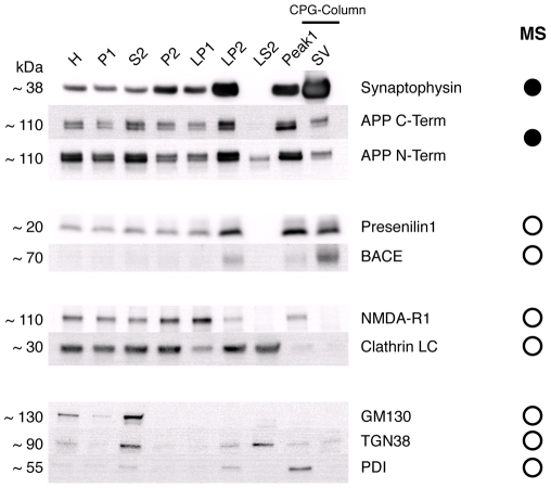
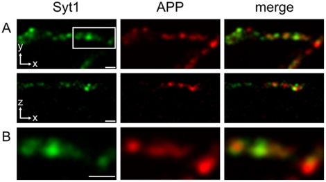
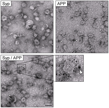
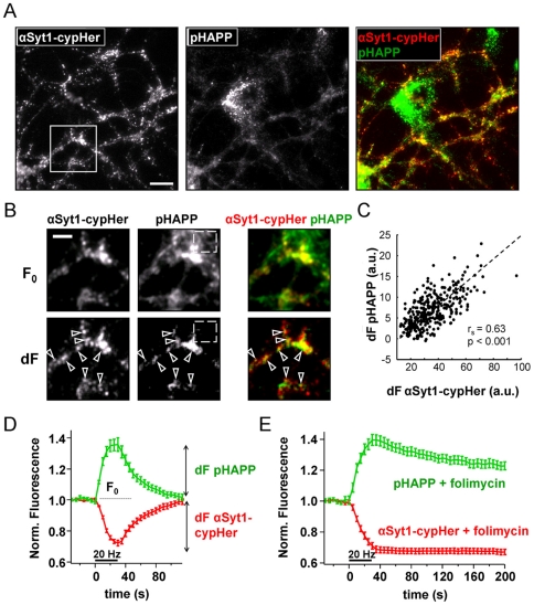
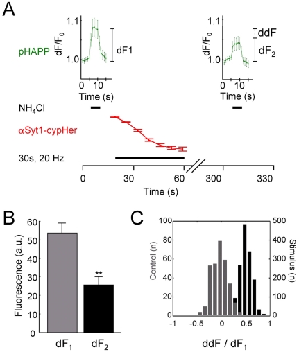
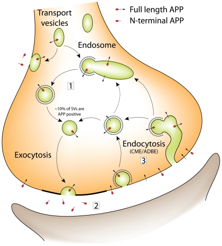
A large body of evidence has implicated amyloid precursor protein (APP) and its proteolytic derivatives as key players in the physiological context of neuronal synaptogenesis and synapse maintenance, as well as in the pathology of Alzheimer's Disease (AD). Although APP processing and release are known to occur in response to neuronal stimulation, the exact mechanism by which APP reaches the neuronal surface is unclear. We now demonstrate that a small but relevant number of synaptic vesicles contain APP, which can be released during neuronal activity, and most likely represent the major exocytic pathway of APP. This novel finding leads us to propose a revised model of presynaptic APP trafficking that reconciles existing knowledge on APP with our present understanding of vesicular release and recycling.


Conflict of interest statement

Competing Interests: The authors have declared that no competing interests exist.

Figures

Figure 1
Figure 1. Endogenous full-length APP localizes to isolated synaptic vesicles.
APP and its associated processing enzymes were found in a preparation of highly pure synaptic vesicles isolated from rat brain by immunoblotting and mass-spectrometry. Fractions were taken during the preparation of synaptic vesicles by a classical subcellular fractionation protocol. The fractions shown represent increasing levels of purification (left to right): whole brain homogenate (H), large cell fragments and nuclei (P1), crude cytosol - small cell fragments including microsomes, small myelin fragments and soluble proteins (S2), isolated nerve terminals ‘synaptosomes’ (P2), nerve terminal plasma membrane (LP1), crude synaptic vesicles (LP2), presynaptic cytosol (LS2), first peak from the size exclusion column containing larger membranes ∼100–200 nm (Peak1) and highly pure synaptic vesicles (SV). 10 or 20 µg of total protein from each of the individual fractions were subjected to SDS-PAGE followed by immunoblotting. Fully glycosylated APP was routinely found in synaptic vesicles by immunoblotting, as were the APP processing enzymes BACE and Presenilin 1. The neuronal specific isoform of clathrin light chain was undetectable. Contamination by ER-Golgi trafficking vesicles was assessed by immunoblotting for GM130 (cis-Golgi), TGN38 (trans-Golgi network) and protein disulfide isomerase (PDI; Endoplasmic Reticulum). The synaptic vesicle fraction was free of contamination by these proteins. The column MS gives the results obtained from mass-spectrometry using the synaptic vesicle fraction. Proteins found in the SV fraction are indicated with filled circles; proteins not found in the SV fraction are indicated with empty circles.
Figure 2
Figure 2. Immunolabeling of fixed hippocampal neurons for APP assessed using 4Pi nanoscopy.
Localization of APP to the presynaptic terminal was initially confirmed by double immunofluorescence labeling of cultured hippocampal neurons. Synaptic boutons were detected by immunostaining for the bona fide synaptic vesicle protein synaptotagmin 1 (Syt1), using an antibody that binds the N-terminal (luminal) domain of the protein. For APP staining, an antibody directed against the C-terminus of the protein was used. Relative protein distributions were assessed using 4Pi nanoscopy. (A) 4Pi nanoscopy images (upper row: xy- projections, lower row: xz-projections) of neuronal processes stained for synaptotagmin and APP. The APP antibody stained punctate structures that were often elongated in shape, presumably representing APP-transport vesicles. However, some puncta also co-labeled synaptic boutons (see B), as identified by the marker synaptotagmin (coverslips N = 9; synaptic boutons n = 113). (B) Detailed images from (A), corresponding to the area marked by the white box. Synaptotagmin positive presynaptic boutons showed a diffuse APP staining throughout most of the vesicle cluster, suggesting that a small proportion of presynaptic APP is localized to synaptic vesicles. Scale bars 1 µm.
Figure 3
Figure 3. APP and synaptophysin co-localize on a fraction of synaptic vesicles.
Synaptic vesicles isolated from rat brain were immunolabeled for synaptophysin (Syp), APP or double labeled (Syp 5 nm gold; APP 10 nm gold), before viewing by negative stain electron microscopy. The low magnification images show the synaptic vesicle preparation to consist solely of small, homogeneously shaped vesicles, with diameters in the range of 40–50 nm. Single labeling resulted in 99% of all vesicles immunopositive for synaptophysin (as previously reported) and 10% of all vesicles immunopositive for APP. Similar results were obtained with double labeling; a double labeled vesicle can clearly be seen in the magnified view. In all images, APP labeling is indicated using arrowheads. For single labeling experiments n = 3; for double labeling experiment n = 2. Scale bars, low magnification 100 nm; high magnification 50 nm.
Figure 4
Figure 4. pHluorin-APP and αSyt1-cypHer co-localize at sites of synaptic activity and accurately report synaptic activity.
A) Unstimulated neurons showed a punctate staining pattern when labeled with either αSyt1-cypHer or pHAPP. pHAPP staining was typically at the detection limit of the system. Overlaying the images showed that pHAPP partly co-localized with αSyt1-cypHer, which also stained untransfected neurons. Note, consistent with the high rate of synthesis and turnover of APP in neurons, pHAPP also seemed to be concentrated in the Golgi compartment of the neuron. B) Comparison of baseline images (F0) and difference images calculated by subtracting the image taken before stimulation from the image taken after stimulation (dF). dF is positive at loci where fluorescence increases upon stimulation (exocytosis) (see also D). As seen in dF images, exocytosis is confined to αSyt1-cypHer positive regions (arrowheads). Images were taken from the region of interest in (A). Stimulation was 30 s, 20 Hz. The boxed region (dashed) in both images illustrates background fluorescence; however, as background fluorescence does not increase upon stimulation it is effectively removed in the difference (dF) image. C) Changes in pHAPP fluorescence (dF) were highly correlated to changes in αSyt1-cypHer fluorescence (arbitrary units). Significance was assessed using a Spearman's rank order test (N = 6; n = 383). D) Time course of fluorescence changes at αSyt1-cypHer positive spots. Synapses labeled with both pHAPP and αSyt1-cypHer show characteristic fluorescence changes upon electrical stimulation (30 s, 20 Hz). Following cessation of the stimulus, fluorescence recovered to pre-stimulus values (average of N = 7 regions, each comprising n>50 boutons; error bars are SEM). E) Folimycin prevented fluorescence recovery at the end of the stimulation. Folimycin is a vacuolar-ATPase inhibitor that blocks the reacidification of synaptic vesicles following endocytosis, proving that pHAPP and αSyt1-cypHer are recovered into synaptic vesicles (average of N = 7 regions, each comprising n>50 boutons; error bars are SEM).
Figure 5
Figure 5. A fraction of pHAPP-fluorescence is lost during electrical stimulation.
A) Schematic diagram detailing the time course experiments used to determine total pHAPP at the synapse. pHAPP content (arbitrary flurescence units) was determined before (dF1) and after electrical stimulation of neurons (dF2), by application of NH4Cl. Note there was a delay between the cessation of stimulation and application of the second ammonium pulse (illustrated by the broken time axis between 60 s–300 s), to allow completion of endocytosis and reuptake of pHAPP. Loss of pHAPP fluorescence (ddF) was calculated as the absolute difference between dF1 and dF2. To minimize photobleaching, image acquisition in the pHAPP channel was limited to the time during ammonium pulses. Periods of NH4Cl addition and electrical stimulation are illustrated by bars on the time axis. αSyt1-cypHer5 fluorescence (arbitrary units) was used as an independent reporter of neuronal activity between the ammonium pulses. Control experiments were performed in an identical fashion, except stimulation was omitted from the protocol. B) Individual boutons show a significant reduction in the absolute NH4Cl-induced fluorescence increase following electrical stimulation (30 s, 20 Hz) (p = 0.0013, paired t-test, N = 4, n = 482; t-test performed on N). C) The absolute difference in fluorescence between the two NH4Cl pulses (ddF) was normalized by expressing as a function of the total pHAPP in the bouton at the start of the experiment (dF1), to take into account slight differences in expression levels from experiment to experiment. Normalized ddFs obtained under control (grey) and stimulated (black) conditions are plotted as population histograms. Values for control experiments are centered on 0, indicating little, or no overall loss of fluorescence from the terminal. In contrast, stimulation resulted in a shift to 0.5, indicative of fluorescence loss.
Figure 6
Figure 6. A revised model for APP trafficking in the presynaptic terminal.
The figure illustrates the various recycling pathways proposed for synaptic vesicles in the presynaptic terminal, and how APP recycling can be integrated. Synaptic vesicle precursors are brought to the presynaptic terminal in transport vesicles. It is thought that these transport vesicles undergo a round of fusion with the plasma membrane followed by retrieval and sorting, possibly in an endosomal intermediate, to form fully functional synaptic vesicles, which are capable of undergoing fusion with the plasma membrane. Following exocytosis, vesicles are retrieved from the plasma membrane by endocytosis. Under physiological conditions this is thought to occur via a clathrin-mediated endocytosis (CME) pathway; it is still unclear whether vesicles lose their coats and are recycled directly, or whether they pass through an endosomal sorting intermediate. Putative endosomes may in fact be formed by activity dependend bulk endocytosis (ADBE) of plasma membrane, which is thought to occur during periods of heavy stimulation. APP trafficking at the synapse can be integrated into our current understanding of synaptic vesicle recycling. It is known that APP is also delivered to the presynaptic terminal in transport vesicles. These transport vesicles either fuse with the plasma membrane, depositing APP on the plasma membrane surface, or alternatively they fuse with an endosomal sorting intermediate (which we postulate is identical to that used during recycling of synaptic vesicles). Hence, synaptic vesicles could incorporate APP when recycling through the endosome (1). During synaptic vesicle exocytosis, APP cleavage products would then be released (2). Slight infidelities in the endocytic process might also mean small amounts of surface-resident APP could be endocytosed, along with bona fide synaptic vesicle proteins. These vesicles may then recycle directly for subsequent rounds of fusion and APP release, or pass through the endosomal system (3). Alternatively APP may be internalized and recycled into vesicles as a result of bulk endocytosis.

References

    1. Sisodia SS, Price DL. Role of the beta-amyloid protein in Alzheimer's disease. FASEB J. 1995;9:366–370. - PubMed
    1. Cirrito JR, Kang JE, Lee J, Stewart FR, Verges DK, et al. Endocytosis is required for synaptic activity-dependent release of amyloid-beta in vivo. Neuron. 2008;58:42–51. - PMC - PubMed
    1. Bell KF, Zheng L, Fahrenholz F, Cuello AC. ADAM-10 over-expression increases cortical synaptogenesis. Neurobiol Aging. 2008;29:554–565. - PubMed
    1. De Strooper B, Saftig P, Craessaerts K, Vanderstichele H, Guhde G, et al. Deficiency of presenilin-1 inhibits the normal cleavage of amyloid precursor protein. Nature. 1998;391:387–390. - PubMed
    1. Cirrito JR, Yamada KA, Finn MB, Sloviter RS, Bales KR, et al. Synaptic activity regulates interstitial fluid amyloid-beta levels in vivo. Neuron. 2005;48:913–922. - PubMed

Publication types

Substances