

Case Reports
doi: 10.3340/jkns.2011.49.3.182.
Epub 2011 Mar 31.
Gas-Filled Intradural Cyst within the Cauda Equine
Affiliations
- PMID: 21556241
- PMCID: PMC3085817
- DOI: 10.3340/jkns.2011.49.3.182
Item in Clipboard
Case Reports
Gas-Filled Intradural Cyst within the Cauda Equine
J Korean Neurosurg Soc.
2011 Mar.
Abstract
A case of radicular pain that resulted from a gas-filled intradural cyst in an 80-year-old male is described. Temporary improvement of radicular pain was observed after CT-guided aspiration. However, recurrent radicular pain led to surgical treatment. In this report, the authors document the radiologic and intraoperative features of a gas-filled intradural cyst that migrated into the nerve root, and propose an optimal treatment plan based on a review of the literature.
Keywords: Cyst; Gas; Intradural; Treatment plan.
Figures

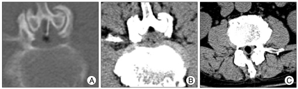
A : Computed tomography scan reveals not only the vacuum phenomena at the L2-3 and L3-4 levels but also intradural gas collection at the L2-3 level. B : Enhanced magnetic resonance image shows a peripheral enhancement of the cyst (arrow).
A : Computed tomography-guided needle aspiration of the intradural gas-filled cyst. The need-le tip is located in the gas-filled cyst. B : There is the remarkable reduction of the cyst and only a sc-anty amount of air in the cyst after the aspiration. C : A month later, axial computed tomography scan shows reaccumulation of the gas-filled intradural cyst.

Intraoperative discography demonstrates the contrast medium flow into the intradural cyst, and partially fills the cyst. Additionally, it shows the air-fluid level in the cyst.

Intraoperative photographs after opening of the dural sac. A : The terminal portion of the intradural nerve root is severely bulging out. B : After removal of the cyst, there is a communication with the L2-3 disc space via a fistula at the ventral dura mater. C : The fistula is sutured with non-absorbable thin nylon thread.

Histological examination reveals multifocal infiltration of chronic inflammatory cells and fibrosis within degenerative fibrocartilage (H & E, ×100).
References
-
- Anda S, Dale LG, Vassal J. Intradural disc herniation with vacuum phenomenon : CT diagnosis. Neuroradiology. 1987;29:407. - PubMed
-
- Bosser V, Dietemann JL, Warter JM, Granel de Solignac M, Beaujeux R, Buchheit F. L5 radicular pain related to lumbar extradural gas-containing pseudocyst. Role of CT-guided aspiration. Neuroradiology. 1990;31:552–553. - PubMed
-
- Demierre B, Ramadan A, Hauser H, Reverdin A, Rilliet B, Bernev J. Radicular compression due to lumbar intraspinal gas pseudocyst: case report. Neurosurgery. 1988;22:731–733. - PubMed
-
- Dillon WP, Kasseff LG, Knackstedt VE, Osborn AG. Computed tomography and differential diagnosis of the extruded lumbar disc. J Comput Assist Tomogr. 1983;7:969–975. - PubMed
-
- Fandino J, Garcia J, Garcia-Abeledo M. Radicular compression by gas in a spinal extra dural cyst. Report on two cases. Neurochirurgie. 1994;40:179–182. - PubMed
Publication types
LinkOut - more resources
Full Text Sources
