Case Reports
. 2011 Mar;49(3):190-3.
doi: 10.3340/jkns.2011.49.3.190. Epub 2011 Mar 31.

Disseminated cysticercosis

Affiliations
Case Reports

Disseminated cysticercosis

Soo Yong Park et al. J Korean Neurosurg Soc. 2011 Mar.

Abstract

Disseminated cysticercosis is a rare form of cysticercosis in which the cysticerci spread out through the whole body. We report the first case of a 39-year-old Mongolian with disseminated cysticercosis. He visited our hospital with generalized tonic-clonic seizure. After extensive investigation from brain computed tomography (CT), spine magnetic resonance imaging (MRI), whole body MRI and pathologic biopsy, he was diagnosed as having cysticercosis involving the brain, subcutaneous tissue, and skeletal muscles through the whole body. We treated him with the albendazole in which case the followed MRI showed that numbers of cystic lesions were copiously decreased. We report an unsual case of disseminated cysticercosis treated with medical therapy.

Keywords: Disseminated cysticercosis; Neurocysticercosis.


Figures

Fig. 1
Fig. 1
Brain CT scan at admission. A : Non-contrast of axial CT scan reveals innumerable cysts with hyperdense "dot" in the brain parenchyma. B : Contrast of axial CT scan demonstrates numerous cysticerci with vesicular stage.
Fig. 2
Fig. 2
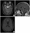
Brain MRI at admission. A : Brain axial, T2-weighted MR image without gadolinium shows that the numerous hyperintense cysts are in brain parenchyma, left optic lesion and temporalis muscle. B : Brain sagittal, post-gadolinium MR image reveals eccentric cyst in the tongue. C : Brain axial, T1-weighted MR image shows innumerable cysts with hyperintense eccentric lesions in the brain parenchyma.
Fig. 3
Fig. 3
Spine MRI at admission. A and B : Spine axial and sagittal T1-weighted MR images without gadolinium show multiple cystic lesions in paraspinal muscle and cysticercus is seen in subarachnoid space on T3 body level. C and D : Spine sagittal, T2-weighted MR images without gadolinium show that numerous cystic lesions are also found in thoracolumbar spine muscle.
Fig. 4
Fig. 4
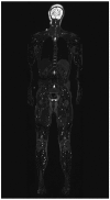
On whole body MRI, T2-weighted image without gadolinium revealing numerous cysticerci on the whole body.
Fig. 5
Fig. 5
Intra-operative finding. A and B : cysticercus has been removed from left temporalis muscle (white arrow). C and D : whitish yellow cystic lesion (black arrow) is seen in subarachnoid space after opening of the dura.
Fig. 6
Fig. 6
Pathologic examination of the mass in the subarachnoid space of the brain (A and B) show the wall of larval cyst with chronic inflammatory cell infiltration.
Fig. 7
Fig. 7
Three months later after treatment, brain axial, T2-weighted MR images scan without gadolinium shows that the numerous cysts are significantly decreased after albendazole treatment. A : Brain sagittal, post-gadolinium MR images also presents decreased number of cystic lesions. B and C : Brain axial, T1-weighted MR image shows disseminated rim enhancing small lesions with mild multifocal edemas in subcortical white matters.

References

    1. Bergsneider M, Holly LT, Lee JH, King WA, Frazee JG. Endoscopic management of cysticercal cysts within the lateral and third ventricles. J Neurosurg. 2000;92:14–23. - PubMed
    1. Bhalla A, Sood A, Sachdev A, Varma V. Disseminated cysticercosis : a case report and review of the literature. J Med Case Reports. 2008:137. - PMC - PubMed
    1. Chang GY, Keane JR. Visual loss in cysticercosis : analysis of 23 patients. Neurology. 2001;57:545–548. - PubMed
    1. Cruz I, Cruz ME, Carrasco F, Horton J. Neurocysticercosis : optimal dose treatment with albendazole. J Neurol Sci. 1995;133:152–154. - PubMed
    1. Del Brutto OH, Rajshekhar V, White AC, Jr, Tsang VC, Nash TE, Takayanagui OM, et al. Proposed diagnostic criteria for neurocysticercosis. Neurology. 2001;57:177–183. - PMC - PubMed

Publication types

LinkOut - more resources