

Imaging signatures of molecular pathology in behavioral variant frontotemporal dementia
- PMID: 21556732
- PMCID: PMC3401589
- DOI: 10.1007/s12031-011-9533-3
Imaging signatures of molecular pathology in behavioral variant frontotemporal dementia
Abstract
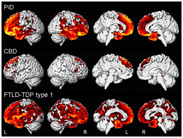
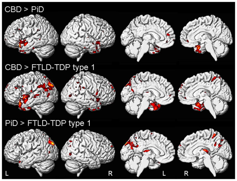
Pathology underlying behavioral variant frontotemporal dementia (bvFTD) is heterogeneous, with the most common pathologies being Pick's disease (PiD), corticobasal degeneration (CBD), and FTLD-TDP type 1. Clinical features are unhelpful in differentiating these pathologies. We aimed to determine whether imaging atrophy patterns differ across these pathologies in bvFTD subjects. We identified 15 bvFTD subjects that had volumetric MRI during life and autopsy: five with PiD, five CBD, and five FTLD-TDP type 1. Voxel-based morphometry was used to assess atrophy patterns in each bvFTD group compared to 20 age- and gender-matched controls. All three pathological groups showed gray matter loss in frontal lobes, although specific patterns of atrophy differed across groups: PiD showed widespread loss in frontal lobes with additional involvement of anterior temporal lobes; CBD showed subtle patterns of loss involving posterior lateral and medial superior frontal lobe; and FTLD-TDP type 1 showed widespread loss in frontal, temporal, and parietal lobes. Greater parietal loss was observed in FTLD-TDP type 1 compared to both other groups, and greater anterior temporal and medial frontal loss was observed in PiD compared to CBD. Imaging patterns of atrophy in bvFTD vary according to pathological diagnosis and may therefore be helpful in predicting these pathologies in bvFTD.
Figures
References
-
- Ashburner J, Friston KJ. Voxel-based morphometry--the methods. NeuroImage. 2000;11:805–821. - PubMed
-
- Ashburner J, Friston KJ. Unified segmentation. NeuroImage. 2005;26:839–851. - PubMed
-
- Bigio EH, Lipton AM, Yen SH, et al. Frontal lobe dementia with novel tauopathy: sporadic multiple system tauopathy with dementia. Journal of neuropathology and experimental neurology. 2001;60:328–341. - PubMed
-
- Boccardi M, Sabattoli F, Laakso MP, et al. Frontotemporal dementia as a neural system disease. Neurobiology of aging. 2005;26:37–44. - PubMed
Publication types
MeSH terms
Substances
Grants and funding
LinkOut - more resources
Full Text Sources
Medical
