. 2011 Nov;216(4):331-45.
doi: 10.1007/s00429-011-0320-2. Epub 2011 May 10.

Projections from the rat pedunculopontine and laterodorsal tegmental nuclei to the anterior thalamus and ventral tegmental area arise from largely separate populations of neurons

Affiliations

Projections from the rat pedunculopontine and laterodorsal tegmental nuclei to the anterior thalamus and ventral tegmental area arise from largely separate populations of neurons

Ericka C Holmstrand et al. Brain Struct Funct. 2011 Nov.

Abstract

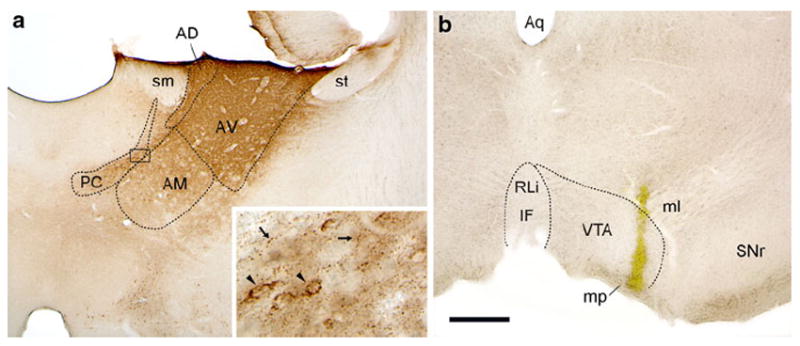
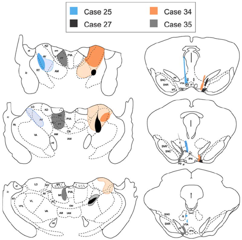
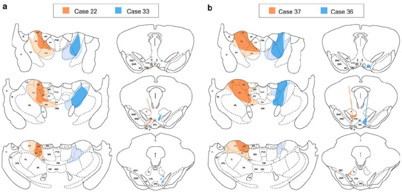
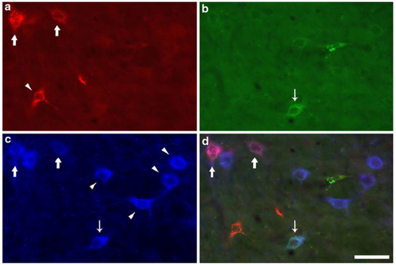
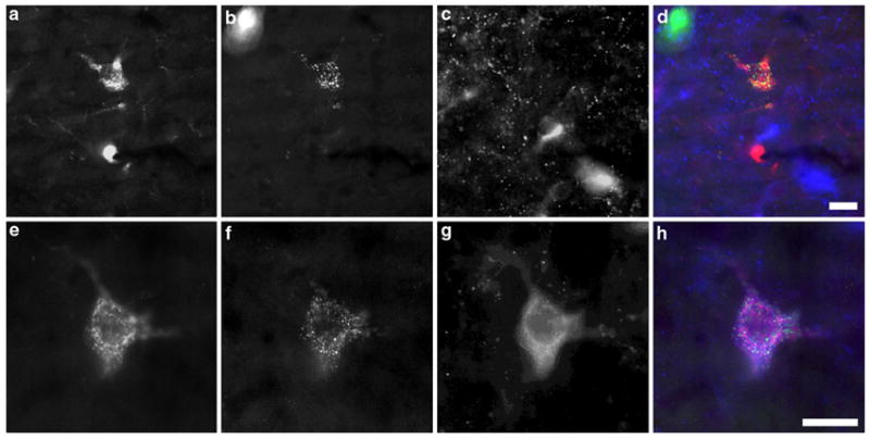
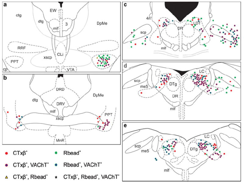
Cholinergic and non-cholinergic neurons in the brainstem pedunculopontine (PPT) and laterodorsal tegmental (LDT) nuclei innervate diverse forebrain structures. The cholinergic neurons within these regions send heavy projections to thalamic nuclei and provide modulatory input as well to midbrain dopamine cells in the ventral tegmental area (VTA). Cholinergic PPT/LDT neurons are known to send collateralized projections to thalamic and non-thalamic targets, and previous studies have shown that many of the afferents to the VTA arise from neurons that also project to midline and intralaminar thalamic nuclei. However, whether cholinergic projections to the VTA and anterior thalamus (AT) are similarly collateralized is unknown. Ultrastructural work from our laboratory has demonstrated that cholinergic axon varicosities in these regions differ both morphologically and with respect to the expression and localization of the high-affinity choline transporter. We therefore hypothesized that the cholinergic innervation to these regions is provided by separate sets of PPT/LDT neurons. Dual retrograde tract-tracing from the AT and VTA indicated that only a small percentage of the total afferent population to either region showed evidence of providing collateralized input to the other target. Cholinergic and non-cholinergic cells displayed a similarly low percentage of collateralization. These results are contrasted to a control case in which retrograde labeling from the midline paratenial thalamic nucleus and the VTA resulted in higher percentages of cholinergic and non-cholinergic dual-tracer labeled cells. Our results indicate that functionally distinct limbic target regions receive primarily segregated signaling from PPT/LDT neurons.


Conflict of interest statement

Conflict of interest The authors declare that they have no conflicts of interest.

Figures

Fig. 1
Fig. 1
Representative CTxβ injection in the anterior thalamus (a) and Rbead injection in the VTA (b) from Case 34. Immunoperoxidase staining for CTxβ shows that the tracer diffused from the center of the injection to fill the AVN. Additional tracer spread was evident in the adjacent AD, AM, and paracentral nucleus (PC). Peroxidase product was also observed in midline and intralaminar nuclei (inset the black box in a indicates the region shown at higher magnification). The punctate appearance of this labeling (small arrows) suggests that it represents anterograde transport. Retrograde labeling of cell soma (arrowheads) was also visible in these areas. The Rbead deposit (b) involved primarily the lateral VTA near the edge of the medial lemniscus (ml) and just above the mammillary peduncle (mp). PC paracentral nucleus, sm stria medullaris, st stria terminalis. Scale bar represents 500 μm in a and b, 50 μm in inset
Fig. 2
Fig. 2
Illustration of the extent of CTxβ injections in the AVN (left panels) and Rbead injections in the VTA (right panels) for three cases in which the thalamic injections were well restricted within the anterior thalamic group (AD, AV, AM and LD), with minimal, or no spread to the midline and intralaminar nuclei (PC, PT, MD, CL, CM). The solid areas show the core of the injections, where CTxβ immunoreactivity was most dense. Hatched regions demarcate the extent of diffusion of CTxβ observed in adjacent regions. An additional control (Case 35) is shown in which the thalamic injection centered on the paratenial nucleus (PT) and did not spread laterally into the AV. Drawings are adapted from Paxinos and Watson (1997)
Fig. 3
Fig. 3
Illustrations depicting CTxβ injections in the AVN (left panels) and Rbead injections in the VTA (right panels) for cases 22 and 33 (a) and cases 36 and 37 (b). As in Fig. 2, the hatched regions show diffusion spread of tracer from the center of the injection represented by the solid regions. Tracer spread was more extensive in these cases, resulting in some involvement of the intralaminar nuclei medial to the AVN. Drawings are adapted from (Paxinos and Watson 1997)
Fig. 4
Fig. 4
Neurons in the PPT that were retrogradely labeled from the AVN or VTA and singly labeled for tracer or dually labeled for tracer and the vesicular acetylcholine transporter (VAChT). Examination of single channel immunofluorescence identified neurons containing: a CTxβ retrogradely transported from the AVN, b Rbeads transported from the VTA, and c VAChT immunoreactivity. d Merger of the three channels shows colocalization of tracers and VAChT in some neurons; thick arrows indicate VAChT-ir neurons projecting to the AVN, whereas the thin arrow identifies a VTA-projecting, VAChT-ir cell. All other neurons are singly labeled (arrowheads). Although some singly labeled VAChT-ir neurons show a faint cellular fluorescence appears in the green channel (b), this was determined to be artifactual by examination at higher magnification (not shown). Scale bar represents 50 μm in all panels
Fig. 5
Fig. 5
Dual tracer labeled cells in the PPT and LDT. Top panels Triple immunofluorescence from the rostral dorsal LDT revealing a non-cholinergic dual-tracer labeled neuron. CTxβ immunoreactivity (a) and Rbeads (b) are both present in a single neuron. VAChT immunoreactivity (c) is present in nearby cells at a different focal plane, but absent from the dual-tracer labeled neuron. Merger of the single fluorescence channels (d) demonstrates colocalization of CTxβ-ir and Rbeads. Bottom panels A triply labeled neuron in the caudal lateral PPT contains CTxβ-ir (e), Rbeads (f), and VAChT immunoreactivity (g). The merged fluorescence channels (h) demonstrate the distinct visual textures of the three markers used, reducing the chance of false positive labeling due to cross-channel bleed-through. Scale bar equals 20 μm in all panels
Fig. 6
Fig. 6
Schematic representation of the spatial distribution of retrogradely labeled neurons within selected sections through the brain-stem tegmentum from Case 33. Panels are presented from most rostral to most caudal (a–f, respectively). CTxβ-ir (red symbols) and Rbead-labeled cells (green symbols) were found at similar rostrocaudal levels throughout the PPT and LDT, and were intermingled within these nuclei on both the ipsilateral (right) and contralateral (left) sides. The subset of afferents that were also VAChT-ir (red or green symbols with blue interiors) was similarly mixed, although cells labeled for both CTxβ and VAChT in this case were observed further rostrally than the Rbead+ VAChT-labeled neurons (as in panel a). For clarity, singly labeled VAChT-ir neurons are not depicted. To conserve space, three additional sections from this case were likewise omitted, but these displayed distributions of cells similar to those shown here

References

    1. Aggleton JP, O’Mara SM, Vann SD, Wright NF, Tsanov M, Erichsen JT. Hippocampal-anterior thalamic pathways for memory: uncovering a network of direct and indirect actions. Eur J Neurosci. 2010;31:2292–2307. - PMC - PubMed
    1. Angelucci A, Clascá F, Sur M. Anterograde axonal tracing with the subunit B of cholera toxin: a highly sensitive immunohistochemical protocol for revealing fine axonal morphology in adult and neonatal brains. J Neurosci Methods. 1996;65:101–112. - PubMed
    1. Barbas H, Henion TH, Dermon CR. Diverse thalamic projections to the prefrontal cortex in the rhesus monkey. J Comp Neurol. 1991;313:65–94. - PubMed
    1. Beckstead RM, Domesick VB, Nauta WJ. Efferent connections of the substantia nigra and ventral tegmental area in the rat. Brain Res. 1979;175:191–217. - PubMed
    1. Beninato M, Spencer RF. A cholinergic projection to the rat substantia nigra from the pedunculopontine nucleus. Brain Res. 1987;412:169–174. - PubMed

Publication types

LinkOut - more resources