. 2011 Aug;39(8):2242-51.
doi: 10.1007/s10439-011-0323-4. Epub 2011 May 10.

Rapid microfluidic perfusion enabling kinetic studies of lipid ion channels in a bilayer lipid membrane chip

Affiliations

Rapid microfluidic perfusion enabling kinetic studies of lipid ion channels in a bilayer lipid membrane chip

Chenren Shao et al. Ann Biomed Eng. 2011 Aug.

Abstract

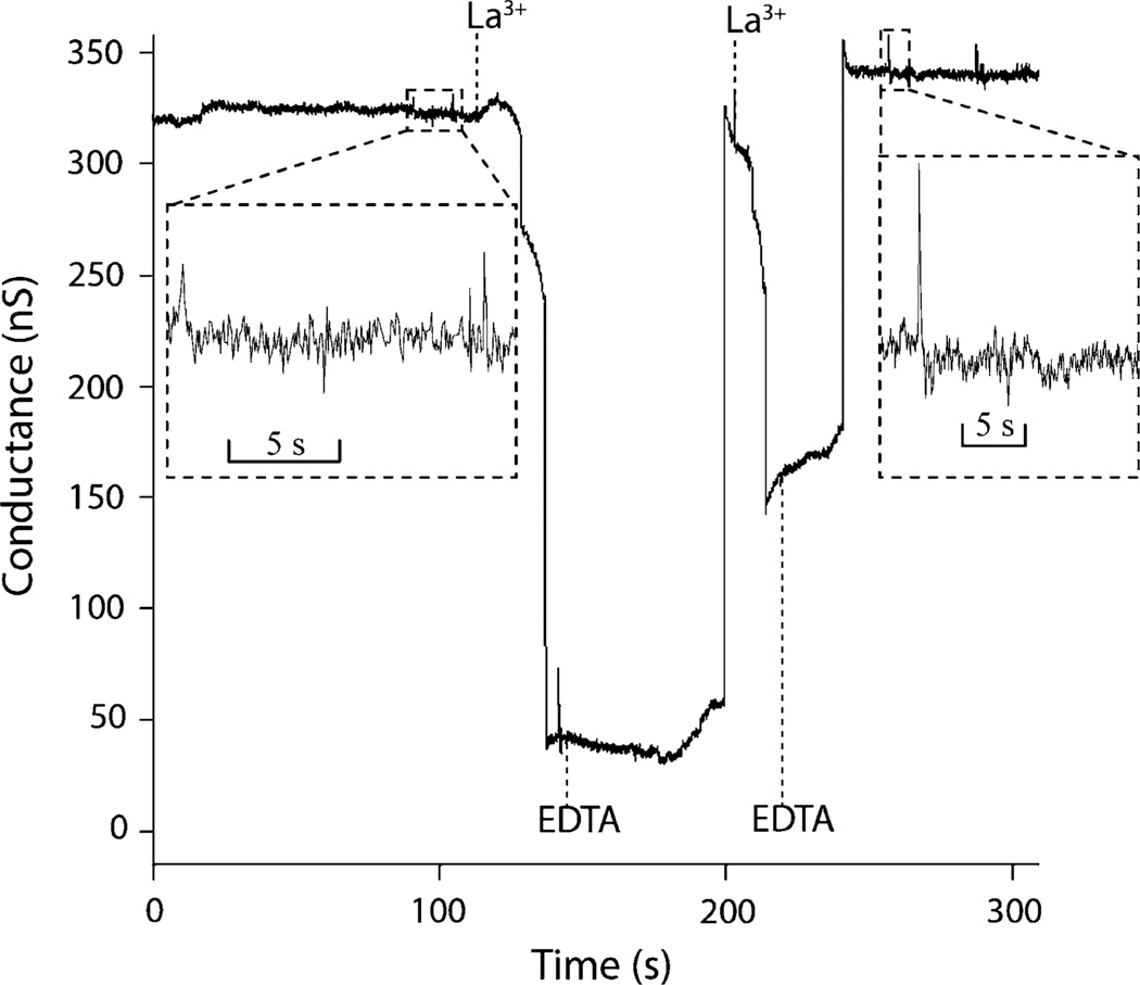
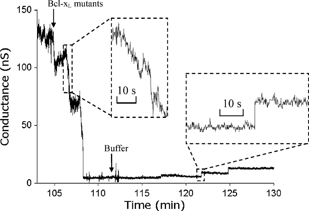
There is growing recognition that lipids play key roles in ion channel physiology, both through the dynamic formation and dissolution of lipid ion channels and by indirect regulation of protein ion channels. Because existing technologies cannot rapidly modulate the local (bio)chemical conditions at artificial bilayer lipid membranes used in ion channel studies, the ability to elucidate the dynamics of these lipid-lipid and lipid-protein interactions has been limited. Here we demonstrate a microfluidic system supporting exceptionally rapid perfusion of reagents to an on-chip bilayer lipid membrane, enabling the responses of lipid ion channels to dynamic changes in membrane boundary conditions to be probed. The thermoplastic microfluidic system allows initial perfusion of reagents to the membrane in less than 1 s, and enables kinetic behaviors with time constants below 10 s to be directly measured. Application of the platform is demonstrated toward kinetic studies of ceramide, a biologically important lipid known to self-assemble into transmembrane ion channels, in response to dynamic treatments of small ions (La(3+)) and proteins (Bcl-x(L) mutant). The results reveal the broader potential of the technology for studies of membrane biophysics, including lipid ion channel dynamics, lipid-protein interactions, and the regulation of protein ion channels by lipid micro domains.


Figures

FIGURE 1
FIGURE 1
(a) An assembled microfluidic perfusion chip. Buffer is introduced through a main channel on the top wafer (inlet 2), with an optional side channel (inlet 1) used to co-inject ion channels or other solutions to the open well. Two perfusion channels (inlets 3 and 4) connect to lower channel beneath the BLM site through a mixer. Two Ag/AgCl electrodes are sealed to chip with adhesive wax. (b) A cross-section schematic view of chip showing the electrical interface used for monitoring transmembrane current. The perfusion pathway (dashed line) passes beneath the BLM site to the waste port.
FIGURE 2
FIGURE 2
Measurements of BLM capacitance over time at different perfusion flow rates up to 3.5 µL/min using a device with a 140 µm diameter membrane aperture. For flow rates up to 2.5 µL/min, negligible increases in membrane capacitance are observed, indicating that the membranes used in this study remain stable over this range. For higher flow rates, capacitance increases linearly with time, corresponding to an observed outward migration of the membrane boundary toward the aperture limits. Regardless of the flow rate, halting perfusion immediately returns the capacitance to its original state provided that the membrane has not been ruptured. At 3.5 µL/min, it takes approximately 5 min to reach the critical rupture point. For chips containing smaller apertures on the order of ~50 µm, maximum continuous pumping flow rates of 10 µL/min are routinely achieved.
FIGURE 3
FIGURE 3
Ceramide channel formation within a membrane pretreated with 50 µM LaCl3 when 50 µM EDTA starts to be perfused to the membrane at time 0. At the given continuous perfusion flow rate (0.2 µL/min), EDTA first reaches the BLM site after an estimated 11.5 min period, followed by a switching time of τ = 72 s to reach the final concentration. Gradual stepwise channel formation is observed beginning 14 min after initiating EDTA perfusion (aperture diameter ~65 µm).
FIGURE 4
FIGURE 4
Dynamic measurements of ceramide channel response to sequential perfusions of 50 µM LaCl3 and 50 µM EDTA. A rapid pulse flow rate of 20 µL/min was used, with a corresponding switching time of τ = 5.8 s (aperture diameter ~60 µm). The time interval between arrival and removal of each injection is estimated as 33.8 s for first LaCl3, 57.9 s for first EDTA, and 16.2 s for second LaCl3. The insets show the sub-second details of ceramide channel activity before and after two cycles of inhibition and recovery.
FIGURE 5
FIGURE 5
A portion of 2-h current recording of ceramide interacting with Y101 Bcl-xL mutants. The ceramide channel is initially stable at ~130 nS before perfusion of Bcl-xL mutants with an estimated switching time of τ = 26.5 s at flow rate of 5 µL/min. The arrival of Bcl-xL mutants (1.75 µg/mL) inhibits the ceramide channel conductance down to nearly zero over a 3 min period. Removal of Bcl-xL from the BLM site by perfusion of buffer leads to slow stepwise recovery of the ceramide channel (aperture diameter ~70 µm). Inset shows a detailed view of stepwise channel disassembly and reassembly.

Similar articles

Cited by

References

    1. Anishkin A, Sukharev S, Colombini M. Searching for the molecular arrangement of transmembrane ceramide channels. Biophys. J. 2006;90:2414–2426. - PMC - PubMed
    1. Antonov VF, Petrov VV, Molnar AA, Predvoditelev DA, Ivanov AS. Appearance of single-ion channels in unmodified lipid bilayer-membranes at the phase-transition temperature. Nature. 1980;283:585–586. - PubMed
    1. Antonov VF, Smirnova EY, Shevchenko EV. Electric-field increases the phase-transition temperature in the bilayer-membrane of phosphatidic-acid. Chem. Phys. Lipids. 1990;52:251–257. - PubMed
    1. Bernardi P, Scorrano L, Colonna R, Petronilli V, Di Lisa F. Mitochondria and cell death—mechanistic aspects and methodological issues. Eur. J. Biochem. 1999;264:687–701. - PubMed
    1. Billen LP, Kokoski CL, Lovell JF, Leber B, Andrews DW. Bcl-XL inhibits membrane permeabilization by competing with Bax. PLoS Biol. 2008;6:e147. - PMC - PubMed

Publication types