

Regional areas and widths of the midsagittal corpus callosum among HIV-infected patients on stable antiretroviral therapies
- PMID: 21556960
- PMCID: PMC4309645
- DOI: 10.1007/s13365-011-0033-6
Regional areas and widths of the midsagittal corpus callosum among HIV-infected patients on stable antiretroviral therapies
Erratum in
- J Neurovirol. 2011 Aug;17(4):380-1. Schifitto, Giavoni [corrected to Schifitto, Giovanni]
Abstract
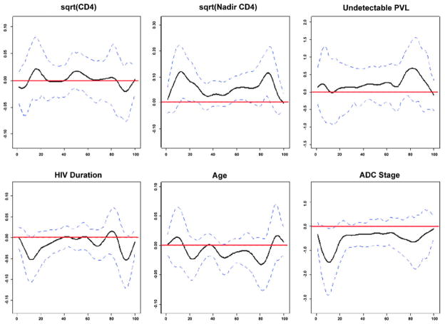
Recent reports suggest that a growing number of human immunodeficiency virus (HIV)-infected persons show signs of persistent cognitive impairment even in the context of combination antiretroviral therapies (cART). The basis for this finding remains poorly understood as there are only a limited number of studies examining the relationship between CNS injury, measures of disease severity, and cognitive function in the setting of stable disease. This study examined the effects of HIV infection on cerebral white matter using quantitative morphometry of the midsagittal corpus callosum (CC) in 216 chronically infected participants from the multisite HIV Neuroimaging Consortium study currently receiving cART and 139 controls. All participants underwent MRI assessment, and HIV-infected subjects also underwent measures of cognitive function and disease severity. The midsagittal slice of the CC was quantified using two semi-automated procedures. Group comparisons were accomplished using ANOVA, and the relationship between CC morphometry and clinical covariates (current CD4, nadir CD4, plasma and CSF HIV RNA, duration of HIV infection, age, and ADC stage) was assessed using linear regression models. HIV-infected patients showed significant reductions in both the area and linear widths for several regions of the CC. Significant relationships were found with ADC stage and nadir CD4 cell count, but no other clinical variables. Despite effective treatment, significant and possibly irreversible structural loss of the white matter persists in the setting of chronic HIV disease. A history of advanced immune suppression is a strong predictor of this complication and suggests that antiretroviral intervention at earlier stages of infection may be warranted.
Figures


References
-
- Ances BM, Ellis RJ. Dementia and neurocognitive disorders due to HIV-1 infection. Semin Neurol. 2007;27:86–92. - PubMed
-
- Anthony IC, Bell JE. The neuropathology of HIV/AIDS. Int Rev Psychiatry. 2008;20:15–24. - PubMed
-
- Antinori A, Arendt G, Becker JT, Brew BJ, Byrd DA, Cherner M, Clifford DB, Cinque P, Epstein LG, Goodkin K, Gisslen M, Grant I, Heaton RK, Joseph J, Marder K, Marra CM, McArthur JC, Nunn M, Price RW, Pulliam L, Robertson KR, Sacktor N, Valcour V, Wojna VE. Updated research nosology for HIV-associated neurocognitive disorders. Neurology. 2007;69:1789–1799. - PMC - PubMed
-
- Archibald SL, Masliah E, Fennema-Notestine C, Marcotte TD, Ellis RJ, McCutchan JA, Heaton RK, Grant I, Mallory M, Miller A, Jernigan TL. Correlation of in vivo neuroimaging abnormalities with postmortem human immunodeficiency virus encephalitis and dendritic loss. Arch Neurol. 2004;61:369–376. - PubMed
-
- Aylward EH, Brettschneider PD, McArthur JC, Harris GJ, Schlaepfer TE, Henderer JD, Barta PE, Tien AY, Pearlson GD. Magnetic resonance imaging measurement of gray matter volume reductions in HIV dementia. Am J Psychiatry. 1995;152:987–994. - PubMed
Publication types
MeSH terms
Substances
Grants and funding
- U24 MH100929/MH/NIMH NIH HHS/United States
- U01 MH083500/MH/NIMH NIH HHS/United States
- 1UO1MH083500/MH/NIMH NIH HHS/United States
- R01MH60565/MH/NIMH NIH HHS/United States
- R01NS35524/NS/NINDS NIH HHS/United States
- U01 AI038855/AI/NIAID NIH HHS/United States
- UL1 TR000124/TR/NCATS NIH HHS/United States
- R01DA15045/DA/NIDA NIH HHS/United States
- K23 MH065857/MH/NIMH NIH HHS/United States
- R01MH65857/MH/NIMH NIH HHS/United States
- K23 MH073416/MH/NIMH NIH HHS/United States
- P30 AI042853/AI/NIAID NIH HHS/United States
- P01 AA019072/AA/NIAAA NIH HHS/United States
- R01NS38841/NS/NINDS NIH HHS/United States
- T32 DA013911/DA/NIDA NIH HHS/United States
- UL1 RR033176/RR/NCRR NIH HHS/United States
- R24 NS038841/NS/NINDS NIH HHS/United States
- T32DA13911/DA/NIDA NIH HHS/United States
- R01AI38855/AI/NIAID NIH HHS/United States
- L30 NS063444/NS/NINDS NIH HHS/United States
- K23-MH073416/MH/NIMH NIH HHS/United States
LinkOut - more resources
Full Text Sources
Medical
Research Materials
