Review
. 2011 Nov-Dec;30(6):1203-24.
doi: 10.1002/mas.20320. Epub 2011 May 9.

Advances in mass spectrometry for the identification of pathogens

Affiliations
Review

Advances in mass spectrometry for the identification of pathogens

Yen-Peng Ho et al. Mass Spectrom Rev. 2011 Nov-Dec.

Abstract

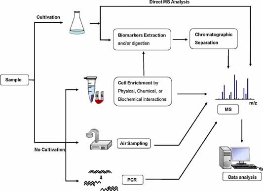
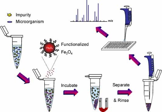
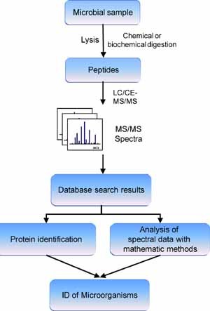
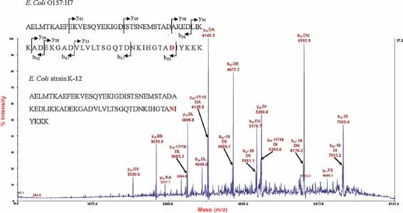
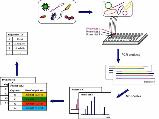
Mass spectrometry (MS) has become an important technique to identify microbial biomarkers. The rapid and accurate MS identification of microorganisms without any extensive pretreatment of samples is now possible. This review summarizes MS methods that are currently utilized in microbial analyses. Affinity methods are effective to clean, enrich, and investigate microorganisms from complex matrices. Functionalized magnetic nanoparticles might concentrate traces of target microorganisms from sample solutions. Therefore, nanoparticle-based techniques have a favorable detection limit. MS coupled with various chromatographic techniques, such as liquid chromatography and capillary electrophoresis, reduces the complexity of microbial biomarkers and yields reliable results. The direct analysis of whole pathogenic microbial cells with matrix-assisted laser desorption/ionization MS without sample separation reveals specific biomarkers for taxonomy, and has the advantages of simplicity, rapidity, and high-throughput measurements. The MS detection of polymerase chain reaction (PCR)-amplified microbial nucleic acids provides an alternative to biomarker analysis. This review will conclude with some current applications of MS in the identification of pathogens.


Figures

Figure 1
Figure 1
Overview of MS‐based approaches in microbial enrichment and identification. [Color figure can be viewed in the online issue, which is available at wileyonlinelibrary.com]
Figure 2
Figure 2
General experimental procedures for microbial enrichment with functionalized magnetic nanoparticles and MS detection. Following the incubation of a microbial solution with functionalized magnetic nanoparticles, microbial cells are isolated and concentrated with a magnet. They are washed. The enriched cells are mixed with a MALDI matrix solution and subjected to MS analysis. [Color figure can be viewed in the online issue, which is available at wileyonlinelibrary.com]
Figure 3
Figure 3
Schematic diagram of a proteomic approach to identify microorganisms based on MS/MS analysis. Microbial proteins are extracted and chemically or biochemically digested. The peptide digests are separated with chromatography and analyzed with MS/MS. The MS/MS spectra are checked against a proteome database to identify the proteins and to deduce the source microorganism. Mathematical algorithms might be applied to evaluate the search results before the microorganisms are identified. [Color figure can be viewed in the online issue, which is available at wileyonlinelibrary.com]
Figure 4
Figure 4
Tandem mass spectrum of a protein marker observed at m/z 7705.6, obtained from the extracted cell lysate of a pathogenic E. coli O157:H7 strain. Many of the fragment ions correspond to backbone cleavage adjacent to aspartic acid (D) and/or glutamic acid (E) residues. The identified protein sequence and the sequence of the non‐pathogenic E. coli K‐12 strain are shown with the spectrum. The two sequences differ by only one amino acid (in bold; aspartic acid vs. asparagine) and, therefore, by only 1 Da in molecular weight. The top‐down proteomics approach can distinguish E. coli O157:H7 from E. coli K‐12. Reprinted and modified with permission from Fagerquist et al. (2010), copyright 2010 American Chemical Society.
Figure 5
Figure 5
PCR‐MS approach‐TIGER: Aliquots of nucleic acids present in a sample are deposited into wells of a micro‐titer plate to begin PCR. Each well contains a pair of broad‐range primers that target a selected domain of microorganisms. PCR products are desalted and electrosprayed into a high‐resolution mass spectrometer to determine their base compositions. The combined base compositions from multiple PCR reactions (multiple primer sets) support the identification of microorganisms in a sample. [Color figure can be viewed in the online issue, which is available at wileyonlinelibrary.com]

References

    1. Abbas‐Hawks C, Voorhees KJ, Miketova P. 2006. Identification of carbohydrate and nucleic acid biomarkers in the pyrolysis electron ionization‐high‐resolution mass spectrum of Brucella neotomae . J Anal Appl Pyrol 76: 6–13.
    1. Adams KL, Steele PT, Bogan MJ, Sadler NM, Martin SI, Martin AN, Frank M. 2008. Reagentless detection of mycobacteria tuberculosis H37RA in respiratory effluents in minutes. Anal Chem 80: 5350–5357. - PubMed
    1. Afonso C, Fenselau C. 2003. Use of bioactive glass slides for matrix‐assisted laser desorption/ionization analysis: Application to microorganisms. Anal Chem 75: 694–697. - PubMed
    1. Al Dahouk S, Nockler K, Scholz HC, Tomaso H, Bogumil R, Neubauer H. 2006. Immunoproteomic characterization of Brucella abortus 1119‐3 preparations used for the serodiagnosis of Brucella infections. J Immunol Methods 309: 34–47. - PubMed
    1. Anhalt JP, Fenselau C. 1975. Identification of bacteria using mass spectrometry. Anal Chem 47: 219–225.

Publication types

MeSH terms

LinkOut - more resources