. 2011 Sep;26(9):2012-25.
doi: 10.1002/jbmr.417.

Genetic evidence points to an osteocalcin-independent influence of osteoblasts on energy metabolism

Affiliations

Genetic evidence points to an osteocalcin-independent influence of osteoblasts on energy metabolism

Yoshihiro Yoshikawa et al. J Bone Miner Res. 2011 Sep.

Abstract

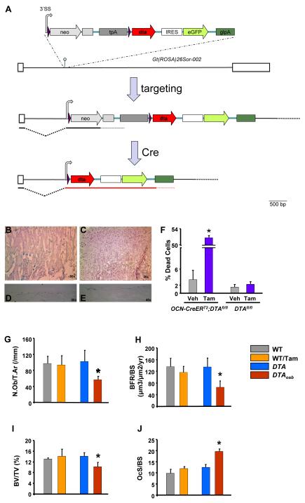
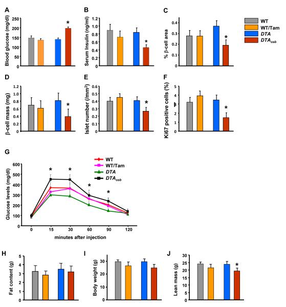
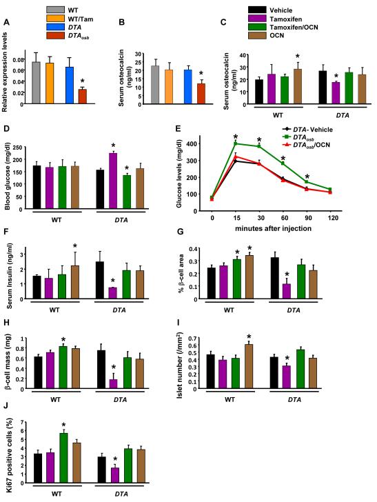
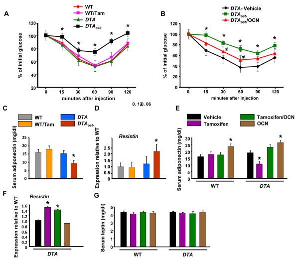
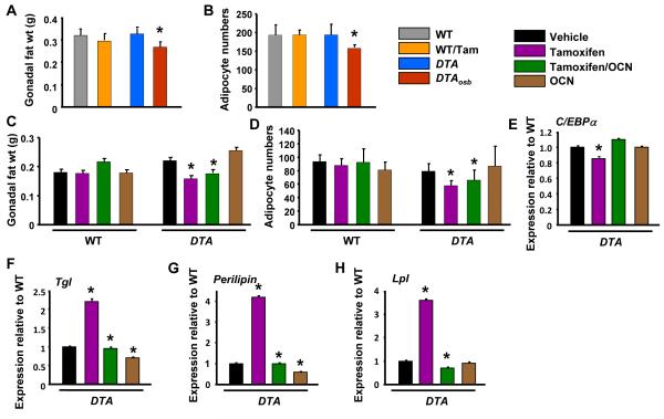
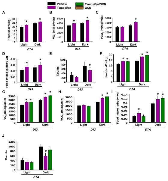
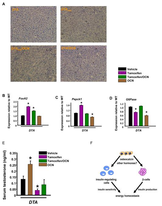
The skeleton has been shown recently to regulate glucose metabolism through an osteoblast-specific hormone, osteocalcin, which favors β-cell proliferation, insulin secretion, insulin sensitivity, and energy expenditure. An implication of this finding is that a decrease in osteoblast numbers would compromise glucose metabolism in an osteocalcin-dependent manner. To test this hypothesis, osteoblasts were inducibly ablated by cross-breeding transgenic mice expressing a tamoxifen-regulated Cre under the control of the osteocalcin promoter with mice in which an inactive form of the diphtheria toxin A chain was introduced into a ubiquitously expressed locus. Ablation of osteoblasts in adult mice profoundly affected glucose metabolism. In a manner similar to what is seen in the case of osteocalcin deficiency, a partial ablation of this cell population resulted in hypoinsulinemia, hyperglycemia, glucose intolerance, and decreased insulin sensitivity. However, and unlike what is seen in osteocalcin-deficient mice, osteoblast ablation also decreased gonadal fat and increased energy expenditure and the expression of resistin, an adipokine proposed to mediate insulin resistance. While administration of osteocalcin reversed (fully) the glucose intolerance and reinstated normal blood glucose and insulin levels, it only partially restored insulin sensitivity and did not affect the improved gonadal fat weight and energy expenditure in osteoblast-depleted mice. These observations not only strengthen the notion that osteoblasts are necessary for glucose homeostasis and energy expenditure but also suggest that in addition to osteocalcin, other osteoblast-derived hormones may contribute to the emerging function of the skeleton as a regulator of energy metabolism.


Figures

Figure 1
Figure 1. Inducible, partial osteoblast ablation in DTAosb mice
(A)The 3′SS-LoxP-EM7-neo-pgkpolyA-tpA-LoxP-dta-ires-eGFP-ßglpA transgene was targeted at the Gt(ROSA)26Sor locus into the XbaI site (hairpin). Only the 002 isoform of Gt(ROSA)26Sor is shown, adapted from Ensembl (release 60 - Nov 2010). Exons are shown as white boxes, whereas native intronic sequences are depicted by a black line. LoxP sites (purple arrows) flank the EM7-neo-pgkpolyA-tpA part of the transgene, enabling its deletion by Cre and concomitant activation of DTA expression. The shared 3′ splice site (3′SS) is shown as a gray curved arrow (hence also marking the direction of transcription. The pgkpolyA immediately following neo is shown as a gray box. The triple polyA (tpA) derived from SV40 is shown as a dark gray box. The intact locus expresses only neo. After exposure to Cre, the floxed region is deleted and expression of DTA is activated. Neo, neomycin phoshotransferase ORF; tpA, triple polyA; DTA, diphtheria toxin A; IRES, internal ribosome entry site; eGPF, enhanced Green Fluorescent Protein; glpA, rabbit beta globin polyA region. (B-J) Eight week-old mice were injected intraperitoneally with vehicle or 0.07mg/g of tamoxifen daily for 10 days. Three days later animals were euthanized and femurs were harvested. Cre expression was examined by assessing β-gal activity in the femurs. For this purpose, whole mount femurs were stained using X-gal, a chromogenic substrate for β-gal. Tissues expressing β-gal are stained with a blue color. Sections shown in the top 2 panels (B and C) were counterstained with eosin whereas sections in panels C and D were not counterstained. All sections were mounted in DPX. Images were acquired with a Nikon 80i Eclipse Microscope using a Retiga digital camera. (B) In the primary spongiosa of femurs obtained from ROSA26-lacZ and OCN-CreERT2 double mutant mice osteoblastic cells expressing β-gal appear blue. (C) There was no staining observed in vehicle-treated double mutant animals. Panels (D) and (E) show two different magnifications of the femoral midshaft in ROSA26-lacZ and OCN-CreERT2 double mutant mice treated with tamoxifen. (F) Calvaria cells were isolated from DTAfl/fl’;OCN-CreERT2 (DTA mice) or from DTAfl/fl (WT mice) control animals. Cells were differentiated in the presence of 50 μM ascorbic acid and β-glycerophosphate for 10 days. Tamoxifen (10−8 M) was added to cultures for 6 hours. Apoptosis was assessed by trypan blue exclusion. Each bar represents individual readings from cells isolated from 6 double mutant and 4 control animals. *p<0.05 vs. vehicle. (G-J) N.Ob/T.Ar, number of osteoblasts per trabecular area; BFR, bone formation rate; BV/TV, bone volume over trabecular volume and OcS/BS, osteoclast surface over bone surface in vertebrae of 2 month-old WT, DTA and DTAosb mice (n=8 mice/group). Bars indicate means ± s.e.m *p<0.05 vs WT and DTA and WT/tamoxifen.
Figure 2
Figure 2. Osteoblast ablation compromises glucoses homeostasis
(A) Blood glucose and (B) serum insulin levels in WT and DTAosb mice at random feeding. (C-E) H&E and insulin staining was used to calculate islet numbers and β-cell area and mass. (F) Ki67 immunostaining showed decreased β-cell proliferation in the pancreas of DTAosb mice. (G) Glucose tolerance test (GTT) in WT and DTAosb mice. (H) total fat content, (I) Body weight, and (J) lean body mass in DTAosb mice. In (J)-(S) n=5 mice/group. In all panels except D and J bars indicate means ± s.e.m *p<0.05 vs WT and DTA and WT/tamoxifen. In D and J *p<0.05 vs WT and DTA. All mice were 2 months of age.
Figure 3
Figure 3. Compromised glucose metabolism and insulin secretion are rescued by Osteocalcin treatment in mice lacking osteoblasts
(A) Real time PCR analysis of osteocalcin expression in bone and (B) and (C) serum osteocalcin levels in DTAosb and osteocalcin-treated DTAosb mice. (D) Blood glucose, and (E) glucose tolerance test in DTAosb mice. (F) Serum insulin levels, (G) Islet numbers, (H) β-cell area, (I) β-cell mass and (J) β-cell proliferation in osteocalcin-treated DTAosb (DTA/Tamoxifen/OCN) mice. In all experiments n=5-8 mice/group. In all panels bars indicate means ± s.e.m *p<0.05 vs WT/vehicle and WT/Tamoxifen and DTA/vehicle. All mice were 2 months of age.
Figure 4
Figure 4. Insulin insensitivity in osteoblast-depleted mice is partially reversed by Osteocalcin
(A) and (B) Insulin tolerance tests (ITT) in WT and DTAosb mice. *p<0.05 vs WT/vehicle and p<0.05 vs DTAosb (C) and (E) Serum adiponectin levels, (D) and (F) real-time PCR analysis of resistin expression in white fat and (G) serum leptin levels in WT and osteocalcin-treated DTAosb (DTA/Tamoxifen/OCN) mice. In (A)-(G) n=5-8 mice/group. In all panels except (B) *p<0.05 vs WT/vehicle and WT/Tamoxifen and DTA/vehicle, In all panels bars indicate means ± s.e.m *p<0.05. All mice were 2 months of age.
Figure 5
Figure 5. Increased fat metabolism in osteoblast-depleted mice is not affected by Osteocalcin
(A) Gonadal fat pad weight and (B) adipocyte numbers in WT and DTAosb mice, n=7 mice/group. (C) Gonadal fat pad weight and (D) adipocyte numbers in osteocalcin-treated WT and DTAosb (DTA/Tamoxifen/OCN) mice, n=7 mice/group. (E-H) Real-time PCR analysis of insulin target genes in white fat of vehicle, tamoxifen or osteocalcin-treated DTA mice, n=7 mice/group. In all panels bars indicate means ± s.e.m *p<0.05 vs WT/vehicle and WT/Tamoxifen and DTA/vehicle. In A p<0.05 vs WT/vehicle and DTA/vehicle. All mice were 2 months of age.
Figure 6
Figure 6. Increased energy expenditure in osteoblast-depleted mice is not affected by Osteocalcin
(A-E) Heat production, oxygen consumption, CO2 expenditure, food intake and total activity (counts) by indirect calorimetric analysis in WT and DTAosb mice, n=4 mice/group. (F-J) Heat production, oxygen consumption, CO2 expenditure, food intake and total activity (counts) by indirect calorimetric analysis in vehicle, tamoxifen or osteocalcin-treated DTA mice, n=4 mice/group. In all panels bars indicate means ± s.e.m *p<0.05 vs WT/vehicle and WT/Tamoxifen and DTA/vehicle. All mice were 2 months of age.
Figure 7
Figure 7. Insulin signaling in the liver of osteoblast-depleted mice is not affected by Osteocalcin
(A) Oil red O staining in liver sections of WT and osteocalcin-treated DTAosb mice. Scale bars are 100 μm. Real-time PCR analysis of (B) FoxA2 expression in the liver of WT and osteocalcin-treated DTAosb (DTA/Tamoxifen/OCN) mice. (C) Pepck1 and (D) G6pase in osteocalcin-treated DTAosb (DTA/Tamoxifen/OCN) mice. In (A)-(L) n=5-8 mice/group. (E) Serum testosterone levels in WT and osteocalcin-treated DTAosb (DTA/Tamoxifen/OCN) mice. In all panels except (A) *p<0.05 vs WT/vehicle and WT/Tamoxifen and DTA/vehicle. All mice were 2 months of age. (F). Osteoblasts act through osteocalcin and perhaps other hormones to affect both the function of β-cells as well as that of other insulin responsive cells in insulin target organs that are sources of glucose production or regulate insulin sensitivity. The total outcome of these interactions is to maintain energy homeostasis.

References

    1. Lee NK, Sowa H, Hinoi E, Ferron M, Ahn JD, Confavreux C, Dacquin R, Mee PJ, McKee MD, Jung DY, Zhang Z, Kim JK, Mauvais-Jarvis F, Ducy P, Karsenty G. Endocrine regulation of energy metabolism by the skeleton. Cell. 2007 Aug 10;130:456–469. - PMC - PubMed
    1. Ferron M, Wei J, Yoshizawa T, Del FA, DePinho RA, Teti A, Ducy P, Karsenty G. Insulin Signaling in Osteoblasts Integrates Bone Remodeling and Energy Metabolism. Cell. 2010 Jul 23;142:296–308. - PMC - PubMed
    1. Fulzele K, Riddle RC, Digirolamo DJ, Cao X, Wan C, Chen D, Faugere MC, Aja S, Hussain MA, Bruning JC, Clemens TL. Insulin Receptor Signaling in Osteoblasts Regulates Postnatal Bone Acquisition and Body Composition. Cell. 2010 Jul 23;142:309–319. - PMC - PubMed
    1. Pittas AG, Harris SS, Eliades M, Stark P, wson-Hughes B. Association between serum osteocalcin and markers of metabolic phenotype. J Clin Endocrinol Metab. 2009 Mar;94:827–832. - PMC - PubMed
    1. Kindblom JM, Ohlsson C, Ljunggren O, Karlsson MK, Tivesten A, Smith U, Mellstrom D. Plasma osteocalcin is inversely related to fat mass and plasma glucose in elderly Swedish men. J Bone Miner Res. 2009 May;24:785–791. - PubMed

Publication types

MeSH terms