

Concise review: Mesenchymal stem cell tumor-homing: detection methods in disease model systems
- PMID: 21557390
- PMCID: PMC4581846
- DOI: 10.1002/stem.645
Concise review: Mesenchymal stem cell tumor-homing: detection methods in disease model systems
Abstract
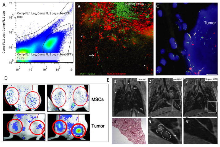
Despite the decline in U.S. cancer incidence and mortality rates, cancer remains the number one cause of death for people under the age of 85 and one in four people in the U.S. will die of cancer, mainly because of metastasis. Recently, interest in mesenchymal stem cell (MSC) tumor-homing has led to inquires into: (a) why MSCs home to tumors, (b) what the inherent protumor and antitumor consequences are, and (c) how to best capitalize on MSC tumor-homing for cell-based diagnostics and therapy. Here, these questions are reviewed and method for addressing them using animal models and tracking methodologies (or, synonymously, detection methodologies) are discussed. First, MSCs in a regenerative and tumor-homing context are reviewed, followed by MSC delivery and genetic labeling methods for tissue model systems. Finally, the use of the nonoptical methods, magnetic resonance imaging, positron emission tomography, and single photon emission computed tomography, along with optical methods, fluorescence imaging and bioluminescent imaging, are reviewed related to tracking MSCs within disease model settings. The benefits and drawbacks of each detection method in animal models is reviewed along with the utility of each for therapeutic use.
Copyright © 2011 AlphaMed Press.
Conflict of interest statement
DISCLOSURE OF POTENTIAL CONFLICTS OF INTEREST
The authors indicate no potential conflicts of interest.
Figures
References
-
- Jemal A, Siegel R, Xu J, Ward E. Cancer Statistics, 2010. [Accessed July 18, 2010];CA: a cancer journal for clinicians. 2010 60(5):277–300. Available at: http://www.ncbi.nlm.nih.gov/pubmed/20610543. - PubMed
-
- Zhang X, Hirai M, Cantero S, et al. Isolation and characterization of mesenchymal stem cells from human umbilical cord blood: Reevaluation of critical factors for successful isolation and high ability to proliferate and differentiate to chondrocytes as compared to mesenchymal stem cells fro. [Accessed February 16, 2011];Journal of cellular biochemistry. 2011 112(4):1206–18. Available at: http://www.ncbi.nlm.nih.gov/pubmed/21312238. - PubMed
-
- Liang L, Dong C, Chen X, et al. Human Umbilical Cord Mesenchymal Stem Cells Ameliorate Mice TNBS-Induced Colitis. [Accessed March 18, 2011];Cell transplantation. 2011 Available at: http://www.ncbi.nlm.nih.gov/pubmed/21396175. - PubMed
-
- Dominici M, Le Blanc K, Mueller I, et al. Minimal criteria for defining multipotent mesenchymal stromal cells. The International Society for Cellular Therapy position statement. Cytotherapy. 2006;8(4):315–317. Available at: http://www.ncbi.nlm.nih.gov/pubmed/16923606. - PubMed
-
- Phinney DG, Prockop DJ. Concise review: mesenchymal stem/multipotent stromal cells: the state of transdifferentiation and modes of tissue repair--current views. [Accessed July 13, 2010];Stem cells (Dayton, Ohio) 2007 25(11):2896–2902. Available at: http://www.ncbi.nlm.nih.gov/pubmed/17901396. - PubMed
Publication types
MeSH terms
Grants and funding
LinkOut - more resources
Full Text Sources
Other Literature Sources
