Review
. 2011 Dec;17(12):2566-84.
doi: 10.1002/ibd.21638. Epub 2011 May 6.

SAMP1/YitFc mouse strain: a spontaneous model of Crohn's disease-like ileitis

Affiliations
Review

SAMP1/YitFc mouse strain: a spontaneous model of Crohn's disease-like ileitis

Theresa T Pizarro et al. Inflamm Bowel Dis. 2011 Dec.

Abstract

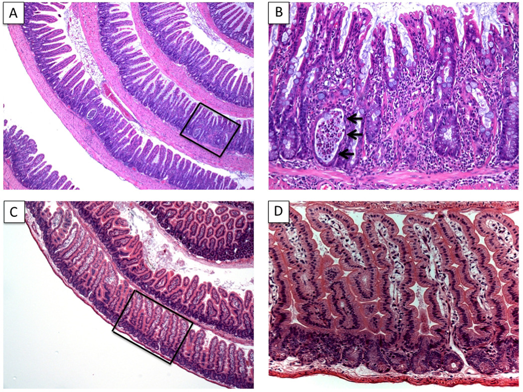
The SAMP1/YitFc mouse strain represents a model of Crohn's disease (CD)-like ileitis that is ideal for investigating the pathogenesis of chronic intestinal inflammation. Different from the vast majority of animal models of colitis, the ileal-specific phenotype characteristic of SAMP1/YitFc mice occurs spontaneously, without genetic, chemical, or immunological manipulation. In addition, SAMP1/YitFc mice possess remarkable similarities to the human condition with regard to disease location, histologic features, incidence of extraintestinal manifestations, and response to conventional therapies. SAMP1/YitFc mice also display a well-defined time course of a predisease state and phases of acute and chronic ileitis. As such, the SAMP1/YitFc model is particularly suitable for elucidating pathways that precede the clinical phenotype that may lead to preventive, and therefore more efficacious, intervention with the natural course of disease, or alternatively, for the development of therapeutic strategies directed against chronic, established ileitis. In this review we summarize important contributions made by our group and others that uncover potential mechanisms in the pathogenesis of CD using this unique murine model of chronic intestinal inflammation.


Figures

Figure 1
Figure 1. Histologic features of SAMP ileitis
Ileal sections from 20 week-old SAMP and AKR control mice were prepared using the Swiss-roll technique. SAMP mice develop spontaneous, transmural inflammation of the terminal ileum characterized by discontinuous inflammatory infiltrates, villous architecture alterations and bowel wall thickening, with hypertrophy of the muscular layers (A, 4X). Higher magnification of panel A (framed area) shows marked villous blunting and distortion, infiltration of acute and chronic immune cells, and the presence of a crypt abscess (arrows) (B, 20X). Uninflamed, control AKR sections are also shown (C, 4X & D, 20X). Panels A and B originally published in Ann N Y Acad Sci, 2009; 1165:301–7.
Figure 2
Figure 2. Mapping of potential chromosomal loci and genes for the susceptibility to SAMP ileitis
Indicated genes include information from both published (27, 42) and preliminary (not yet confirmed) data. Many of the depicted genes are associated with epithelial barrier as well as immune regulatory functions. Portions of the figure originally published in Gastroenterol, 2003; 125:477–90.
Figure 3
Figure 3. Morphologic and functional epithelial alterations in SAMP mice
Epithelial hyperplasia, primarily of cells derived from secretory cell lineages, including Paneth (A & B, 40X) and goblet (C & D, PAS staining, 20X) cells, as well as an altered villous:crypt ratio is observed in the ileum of SAMP (A & C) compared to AKR control mice (B & D). Representative semi-thin sections (□200 nm) of ileal crypt region in SAMP mice (E, 100X) demonstrate abnormal Paneth cell number (arrowheads), and presence of “intermediate cells” (Paneth/goblet hybrid cells, arrows) compared to AKR mice (F, 100X). Scanning electron micrographs show severe villous expansion and blunting, as well as abundant mucus production in SAMP (G) versus AKR controls (H). (A–H, originally published in Ann N Y Acad Sci, 2009; 1165:301–7). (I) Time course of SAMP ileitis versus AKR controls measured by total inflammatory scores (left panel), compared to small intestinal permeability, measured in vivo by Fractional Excretion (FE) of regional-specific sugar probes (lactulose:mannitol) (right panel) demonstrate that small intestinal paracellular permeability is impaired prior to the onset of ileitis in SAMP mice. (J) Early, pre-inflammatory alterations in small intestinal barrier function are present in the absence of the enteric microflora. Small intestinal permeability and ileal inflammation were measured in 3- and 20-week-old GF SAMP mice. N≥4 per group; *P<0.05 vs. age-matched AKR and SPF SAMP. Data originally published in J Exp Med, 2006; 203:541–52 and in Ann N Y Acad Sci, 2009; 1165:301–7.
Figure 4
Figure 4. Summary of the epithelial contribution to SAMP ileitis
Schematic representation of IEC biology from healthy, uninflamed gut and from SAMP ileitis. Several molecular aberrations in SAMP IEC may exist in initiating and sustaining chronic intestinal inflammation characteristic of these mice. A primary dysregulation of TJ protein expression is characterized by marked upregulation of CLDN2 and downregulation of OCCLDN, leading to increased epithelial paracellular permeability and penetration of luminal antigens into the underlying gut mucosa. During health, probiotics have the ability to augment epithelial-derived TNF, which is pivotal for maintaining epithelial barrier function and innate immune defenses; however, this mechanism is dysregulated in SAMP mice. Increased levels of epithelial-derived pro-inflammatory mediators, including IL-33, CRS4C, STAT3 and RELM®, and a decrease in anti-inflammatory molecules, such as PPARγ, have the ability to initiate and activate inflammatory cascades leading to chronic ileitis. ZO-1, -2, zonula occludens-1, -2; TNF, tumor necrosis factor: IL-6, IL-6R, interleukin-6, IL-6 receptor; IL-33, interleukin-33; CRS4C, cysteine-rich sequence 4C; STAT-3, signal transducer and activator of transcription-3; RELM®, resistin-like molecule-®; PPARγ, peroxisome proliferator-activated receptor-γ.
Figure 5
Figure 5. Epithelial-DC morphology is significantly altered in SAMP mice
Massive recruitment of CD11c+ DCs and villous expansion in SAMP (left panel) ileum vs. AKR (right panel) mice occurs early during the onset of inflammation, and increases as disease progresses. 40 week-old SAMP and AKR ilea were fixed in 1% PFA solution and embedded in OCT. Frozen sections were stained for CD11c (green) and phalloidin (blue), and images acquired using a Leica SP5 confocal microscope and analyzed by Bitplane Imaris.
Figure 6
Figure 6. Mixed Th1/Th2 cytokine phenotype characteristic of SAMP mice
Cytokine profiles from gut-associated lymphoid tissues of SAMP mice change from a Th1 predominant (early/inductive phase) to a mixed Th1/Th2 (chronic phase) phenotype as ileitis severity progresses. (A) Total RNA was extracted from SAMP at different ages. Expression of cytokine mRNA levels were measure by real-time PCR and normalized to the 18S ribosomal RNA. All values are expressed relative to the mean value from the <5-wk group, and the y-axis indicates relative mRNA expression. Total inflammatory index for each time point is also indicated; *P<0.05 and **P<0.01. (B) Cytokine production from anti-CD3 stimulated LPL from SAMP vs. AKR ilea. (C) Comparative study of ratios of IFN-γ/IL-4 and IFN-γ/IL-5 secretion by ileal vs. cecal LPL from SAMP mice. Cytokine levels were measured by Cytometric Bead Array and IL-13 by ELISA. Data are expressed as mean±SEM. Data was originally published in Gastroenterol, 2005; 128:654–66.
Figure 7
Figure 7. Impact of bacterial flora on the development of SAMP ileitis
(A) Distribution of ileitis severity in GF vs. SPF SAMP mice (>13 wks of age); TIS, total inflammatory score. (B) Total RNA was extracted from the ileum of GF and SPF SAMP, and mRNA quantified by real-time PCR and normalized to 18S ribosomal RNA. GATA3:T-bet ratio represents the ratio of GATA3 and T-bet mRNA after normalization. Data are expressed as mean±SEM. (C) Activated (anti-CD3/anti-CD28) splenocytes from SPF SAMP cultured in the presence/absence of fecal antigens (Fag). IL-4 or IFNγ were produced by CD4+ lymphocytes, detected by intracellular staining. (D) Reactivity of mucosal lymphocytes from inflamed SPF SAMP mice to stimulation with fecal antigens (FA) prepared from GF or SPF mice. CD4+ MLN T cells represent the effector population. Syngeneic splenocytes were pulsed with FA overnight, irradiated and used as antigen-presenting cells (APC) as indicated. Proliferation of effector lymphocytes was estimated at 72h by measuring [3H]thymidine incorporation for the last 18h of culture. Data originally published in J Immunol, 2007; 178:1809–18. Copyright 2007. The American Association of Immunologists, Inc.
Figure 8
Figure 8. Summary of response to therapies in the SAMP model of CD-like ileitis
Efficacy of different therapies to treat SAMP ileitis before the onset of chronic intestinal inflammation (pre-treatment), during established disease (treatment), or using the adoptive transfer model utilizing SAMP MLN cells transferred into naïve SCID recipients. Bars represent total inflammatory scores of various treatment modalities expressed as the percentage of placebo-treated SAMP (set as 100%) (i.e., ileal inflammation in SAMP mice was decreased by 40% following a pretreatment strategy of metronidazole + ciprofloxacine). NS, not significant.

References

    1. Peyrin-Biroulet L, Loftus EV, Jr, Colombel JF, et al. The natural history of adult Crohn's disease in population-based cohorts. Am J Gastroenterol. 2010;105:289–297. - PubMed
    1. Hugot JP, Chamaillard M, Zouali H, et al. Association of NOD2 leucine-rich repeat variants with susceptibility to Crohn's disease. Nature. 2001;411:599–603. - PubMed
    1. Ogura Y, Bonen DK, Inohara N, et al. A frameshift mutation in NOD2 associated with susceptibility to Crohn's disease. Nature. 2001;411:603–606. - PubMed
    1. Peltekova VD, Wintle RF, Rubin LA, et al. Functional variants of OCTN cation transporter genes are associated with Crohn disease. Nat Genet. 2004;36:471–475. - PubMed
    1. Stoll M, Corneliussen B, Costello CM, et al. Genetic variation in DLG5 is associated with inflammatory bowel disease. Nat Genet. 2004;36:476–480. - PubMed

Publication types