Review
. 2011 Jun;71(6):403-18.
doi: 10.1002/dneu.20818.

Hooks and comets: The story of microtubule polarity orientation in the neuron

Affiliations
Review

Hooks and comets: The story of microtubule polarity orientation in the neuron

Peter W Baas et al. Dev Neurobiol. 2011 Jun.

Abstract

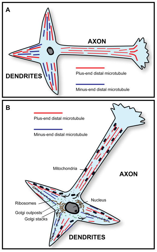
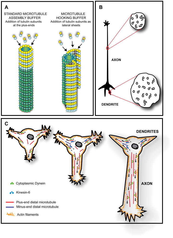
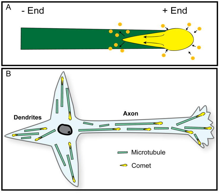
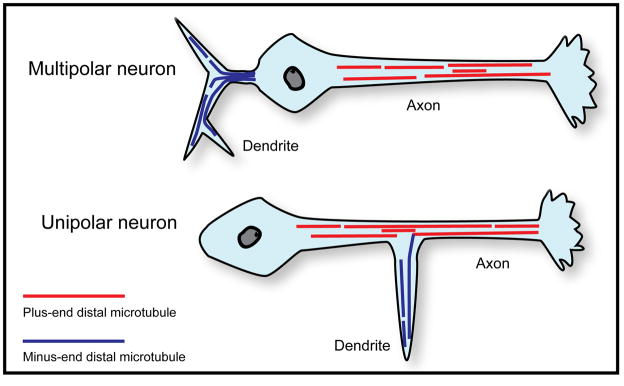
It is widely believed that signature patterns of microtubule polarity orientation within axons and dendrites underlie compositional and morphological differences that distinguish these neuronal processes from one another. Axons of vertebrate neurons display uniformly plus-end-distal microtubules, whereas their dendrites display non-uniformly oriented microtubules. Recent studies on insect neurons suggest that it is the minus-end-distal microtubules that are the critical feature of the dendritic microtubule array, whether or not they are accompanied by plus-end-distal microtubules. Discussed in this article are the history of these findings, their implications for the regulation of neuronal polarity across the animal kingdom, and potential mechanisms by which neurons establish the distinct microtubule polarity patterns that define axons and dendrites.


Figures

Figure 1
Figure 1
A, Microtubule polarity patterns in typical vertebrate axons and dendrites. Axons contain uniformly oriented microtubules (plus-end-distal), while dendrites contain non-uniformly oriented microtubules. B, Microtubule polarity patterns as the basis for asymmetric organelle distribution in vertebrate neurons. A simple yet attractive model for the asymmetric distribution of organelles in the vertebrate neuron posits that axons and dendrites contain those organelles that are transported from the cell body toward plus ends of microtubules (such as mitochondria) but only dendrites contain those organelles that are transported from the cell body toward minus ends of microtubules (such as Golgi outposts and ribosomes). Arrows in B show the direction of organelle movement.
Figure 2
Figure 2
A and B, The hooking procedure for ascertaining microtubule polarity orientation. In this technique, microtubule assembly is allowed to occur in the presence of a rather unique microtubule assembly buffer that promotes tubulin subunit addition preferentially along the sides of existing microtubules rather than at their ends. A, The newly added tubulin forms a protofilament sheet that curves around the existing microtubule. If the reaction is stopped early enough, the curved sheet stops short of closing, and appears, if viewed in cross-section, as a hooked appendage on the microtubule. B, A clockwise hook corresponds to the plus end of the microtubule facing the observer, while a counterclockwise hook corresponds to the minus end of the microtubule facing the observer. Thus, in cross-sections of axons, virtually all hooks are oriented in the same direction, while in the case of dendrites, there are clockwise hooks on some microtubules and counterclockwise hooks on other microtubules. In using this approach to assess microtubule polarity orientation in cells, it is necessary to include a detergent in the hooking mixture in order for the exogenous tubulin subunits to enter the cell. C, Model for establishing and maintaining microtubule polarity patterns in vertebrate axons and dendrites based on cytoplasmic dynein and kinesin-6. It has been proposed that cytoplasmic dynein drives microtubules from the cell body into developing axons and dendrites with plus-end-leading. Then, kinesin-6 transports microtubules with minus-ends-leading specifically into the developing dendrites but not into the developing axon. Axonal microtubule polarity orientation thus remains uniformly plus-end-distal, while dendritic microtubule polarity orientation becomes non-uniform (i.e., mixed). The first neuron shown in the figure is a hippocampal neuron with multiple immature processes. The middle neuron shows the next stage of development wherein one of the immature processes has begun to differentiate into the axon. The third neuron shows later yet in development when the axon has further elongated and the remaining immature processes have become dendrites. As shown schematically, cytoplasmic dynein can transport microtubules with their plus-ends-leading either against other (longer) microtubules or against the actin cytoskeleton, whereas kinesin-6 transports microtubules with their minus-ends-leading exclusively against plus-end-distal microtubules.
Figure 3
Figure 3. The +tip approach for ascertaining microtubule polarity orientation
This technique takes advantages of the properties of +tips, such as EB1 or EB3, which associate with the plus ends of microtubules as they assemble. The association is transient and exists only in the vicinity of the plus end. Therefore, as the microtubule continues to assemble, the +tip molecules lose association with the older region of the microtubule while continuing to associate with the plus end. A, When GFP fusions of +tips are expressed in cells, they appear as comet-shaped fluorescence at the plus ends of the microtubules. The greatest intensity of fluorescence is toward the plus end of the microtubule, while the comet tail (representing the gradual loss of +tip association with the microtubule) is directed toward the minus end of the microtubule (see arrows). B, Uniform and non-uniform microtubule polarity patterns in vertebrate neurons as revealed by the +tip “comet” approach.
Figure 4
Figure 4. Microtubule polarity patterns in fly axons and dendrites
Drosophila neurons can be multipolar, with an axon and dendrites extending from the cell body, or unipolar, with dendrites extending out of the axon. Axons of Drosophila neurons contain uniformly plus-end-distal microtubules, while the dendrites of these neurons contain nearly uniform (90–95%) minus-end-distal microtubules.

References

    1. Ahmad FJ, Echeverri CJ, Vallee RB, Baas PW. Cytoplasmic dynein and dynactin are required for the transport of microtubules into the axon. J Cell Biol. 1998;140:391–401. - PMC - PubMed
    1. Ahmad FJ, He Y, Myers KA, Hasaka TP, Francis F, Black MM, Baas PW. Effects of dynactin disruption and dynein depletion on axonal microtubules. Traffic. 2006;7:524–537. - PubMed
    1. Akhmanova A, Steinmetz MO. Tracking the ends: a dynamic protein network controls the fate of microtubule tips. Nat Rev Mol Cell Biol. 2008;9:309–322. - PubMed
    1. Alberts B. Molecular biology of the cell. Vol. 16. New York: Garland Science; 2008. pp. 965–1052.
    1. Baas PW, Ahmad FJ. The plus ends of stable microtubules are the exclusive nucleating structures for microtubules in the axon. J Cell Biol. 1992;116:1231–1241. - PMC - PubMed

Publication types