Review
. 2011 Dec;71(12):1212-26.
doi: 10.1002/dneu.20905.

The retinal mosaics of opsin expression in invertebrates and vertebrates

Affiliations
Review

The retinal mosaics of opsin expression in invertebrates and vertebrates

Jens Rister et al. Dev Neurobiol. 2011 Dec.

Abstract

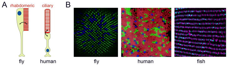
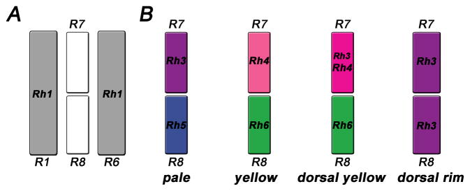
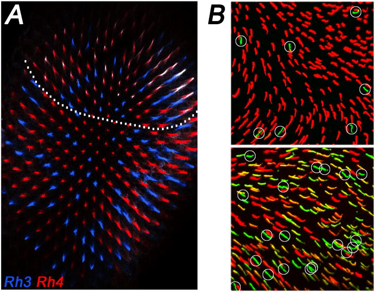
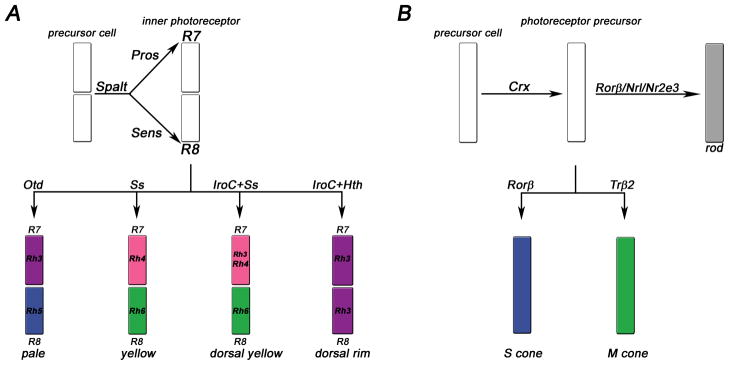
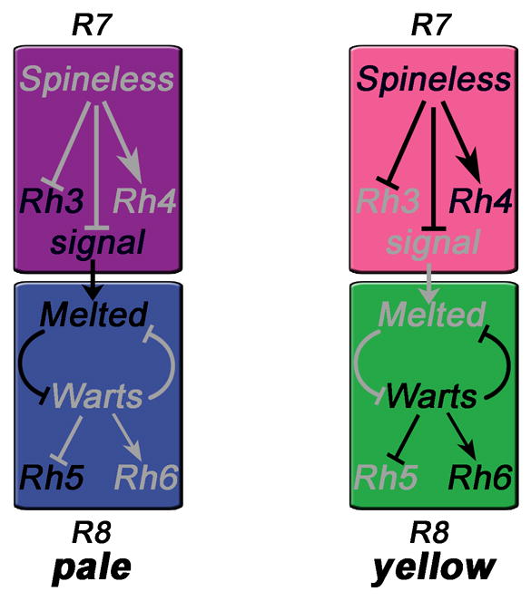
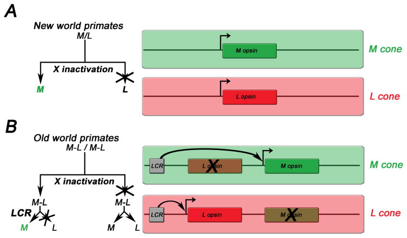
Color vision is found in many invertebrate and vertebrate species. It is the ability to discriminate objects based on the wavelength of emitted light independent of intensity. As it requires the comparison of at least two photoreceptor types with different spectral sensitivities, this process is often mediated by a mosaic made of several photoreceptor types. In this review, we summarize the current knowledge about the formation of retinal mosaics and the regulation of photopigment (opsin) expression in the fly, mouse, and human retina. Despite distinct evolutionary origins, as well as major differences in morphology and phototransduction machineries, there are significant similarities in the stepwise cell-fate decisions that lead from progenitor cells to terminally differentiated photoreceptors that express a particular opsin. Common themes include (i) the use of binary transcriptional switches that distinguish classes of photoreceptors, (ii) the use of gradients of signaling molecules for regional specializations, (iii) stochastic choices that pattern the retina, and (iv) the use of permissive factors with multiple roles in different photoreceptor types.


Figures

Figure 1
Figure 1
Photoreceptor morphology and retinal mosaics in invertebrates and vertebrates. A) Light is absorbed by specialized photoreceptor compartments (red), called rhabdomeres in invertebrates and discs in vertebrates. Adapted from (Arshavsky, 2003) with permission from AAAS. B) Stochastic (fly, human) or highly organized (fish) opsin expression in retinal mosaics. The fly R8 photoreceptors express either the short wavelength Rh5 (blue) or the medium wavelength sensitive Rh6 (green). Humans have three types of cones for short (blue), medium (green) and long wavelengths (red). Adapted from (Roorda and Williams, 1999). The zebrafish retina differs from the previous two mosaics, as it has a precisely arranged distribution of cones (UV and blue; adapted from (Allison et al., 2010)).
Figure 2
Figure 2
(A) ‘Outer’ photoreceptors (grey, only R1 and R6 are shown) express Rh1 and surround the ‘inner’ R7/8, which occur in four subtypes (B) that express different rhodopsins: pale (Rh3/Rh5), yellow (Rh4/Rh6), dorsal yellow (Rh3+Rh4/Rh6) and dorsal rim (Rh3/Rh3). Note that dorsal rim R7/8 are monochromatic and have enlarged rhabdomeres.
Figure 3
Figure 3
Regional differences in opsin expression. (A) Fly dorsal yellow ommatidia coexpress (white) Rh3 (blue) and Rh4 (red). Note that these specialized ommatidia occur only in the dorsal third of the eye (indicated by dotted line). (B) The dorsal mouse retina (top) has mostly M opsin (red) and few S opsin (green, circled) positive cones. In the ventral retina (bottom), the majority of cones show variable levels of coexpression of M and S opsin. Cones that appear to predominantly express S opsin (green, circled) are much more frequent than in the dorsal part of the retina. Figure adapted from (Haverkamp et al., 2005).
Figure 4
Figure 4
Simplified schematic of major cell fate decisions that create the retinal mosaic in the fly (A) and mouse (B) eyes. A) In flies, Spalt specifies inner photoreceptor fate. Prospero (Pros) determines R7 fate and Senseless (Sens) R8 fate. Then, a choice is made for one of the four R7/8 subtypes. B) In the mouse retina, Crx is expressed in all photoreceptor precursor cells. Rods are distinguished from cones by the expression of Rorβ, Nrl and Nr2e3. Cones adopt either short (S) or medium (M) cone fate depending on the expression of either Rorβ or Trβ2, respectively. Note that Rorβ plays a dual role in the rod and S cone pathways. For details, see text.
Figure 5
Figure 5
Pale vs. yellow cell fate decisions in R7 and instruction of R8 (Mikeladze-Dvali et al., 2005). Expression of the transcription factor Spineless in a subset of R7 cells induces yellow fate and Rh4 expression (right). The other R7s express Rh3 by default (left) and send a signal to the underlying R8, which appears to be received by Melted and leads to Rh5 expression. In yellow R8s, the signal is absent and Warts represses Melted as well as Rh5, promoting the default Rh6 fate. Grey indicates repressed genes and inactive regulatory pathways.
Figure 6
Figure 6
Cell-fate decisions (left) and opsin configurations (right) underlying trichromacy in new world (A) and old world primates (B). A) In new world primates (e.g. squirrel monkeys), X chromosomal inactivation leads to expression of each of two different alleles of an M/L opsin (green/red), in addition to an S opsin (not shown) in heteroallelic females that are therefore trichromats. Males and homoallelic females are dichromats. B) In old world primates (e.g. humans), X-chromosomal inactivation and a stochastic choice by a locus control region (LCR) ensure the exclusive expression of either an L or an M opsin that are arranged in a tandem on the X-chromosome. This leads to trichromacy in both males and females. The decision trees to the left exemplify how the choice for M is made in the two different contexts.

Similar articles

Cited by

References

    1. Ahnelt PK, Kolb H. The mammalian photoreceptor mosaic-adaptive design. Prog Retin Eye Res. 2000;19:711–777. - PubMed
    1. Akhmedov NB, Piriev NI, Chang B, Rapoport AL, Hawes NL, Nishina PM, Nusinowitz S, Heckenlively JR, Roderick TH, Kozak CA, Danciger M, Davisson MT, Farber DB. A deletion in a photoreceptor-specific nuclear receptor mRNA causes retinal degeneration in the rd7 mouse. Proc Natl Acad Sci U S A. 2000;97:5551–5556. - PMC - PubMed
    1. Allison WT, Barthel LK, Skebo KM, Takechi M, Kawamura S, Raymond PA. Ontogeny of cone photoreceptor mosaics in zebrafish. J Comp Neurol. 2010;518:4182–4195. - PMC - PubMed
    1. Applebury ML, Antoch MP, Baxter LC, Chun LL, Falk JD, Farhangfar F, Kage K, Krzystolik MG, Lyass LA, Robbins JT. The murine cone photoreceptor: a single cone type expresses both S and M opsins with retinal spatial patterning. Neuron. 2000;27:513–523. - PubMed
    1. Applebury ML, Farhangfar F, Glosmann M, Hashimoto K, Kage K, Robbins JT, Shibusawa N, Wondisford FE, Zhang H. Transient expression of thyroid hormone nuclear receptor TRbeta2 sets S opsin patterning during cone photoreceptor genesis. Dev Dyn. 2007;236:1203–1212. - PubMed

Publication types