. 2011 Sep;71(9):747-58.
doi: 10.1002/dneu.20907.

The actin nucleating Arp2/3 complex contributes to the formation of axonal filopodia and branches through the regulation of actin patch precursors to filopodia

Affiliations

The actin nucleating Arp2/3 complex contributes to the formation of axonal filopodia and branches through the regulation of actin patch precursors to filopodia

Mirela Spillane et al. Dev Neurobiol. 2011 Sep.

Abstract

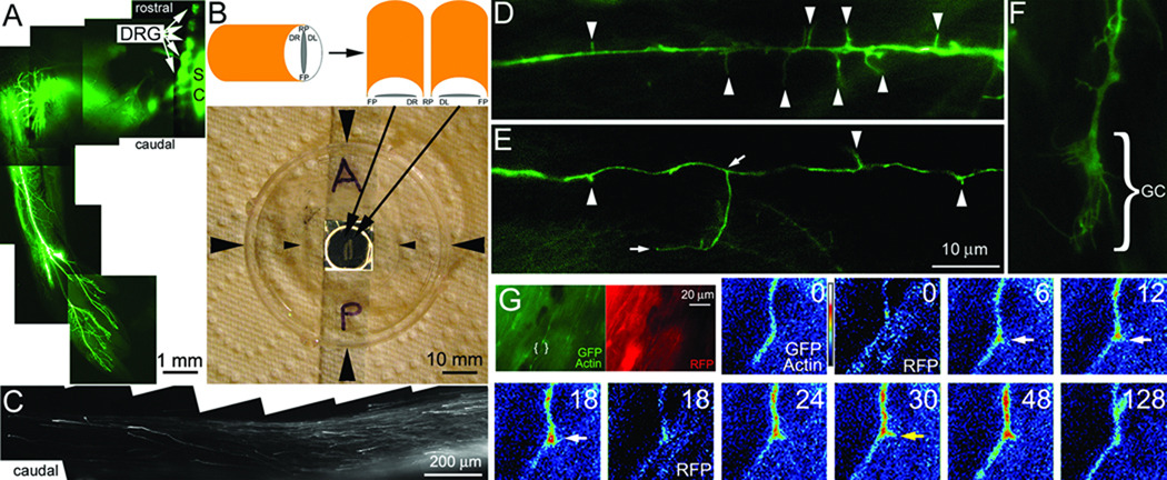
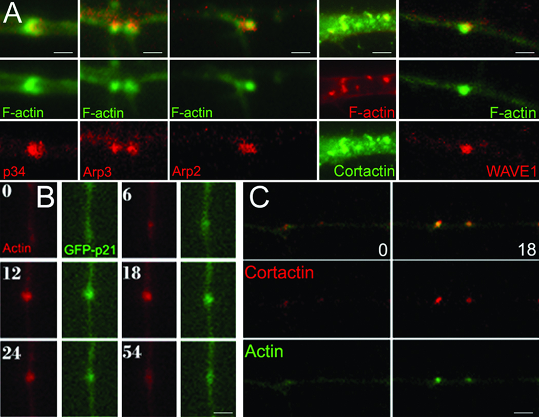
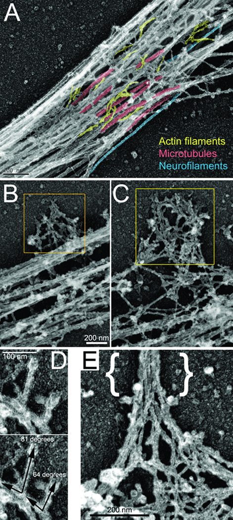
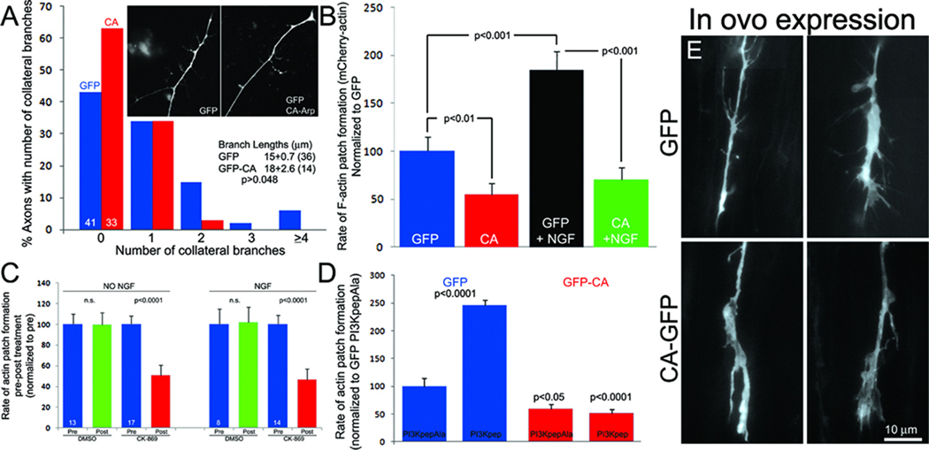
The emergence of axonal filopodia is the first step in the formation of axon collateral branches. In vitro, axonal filopodia emerge from precursor cytoskeletal structures termed actin patches. However, nothing is known about the cytoskeletal dynamics of the axon leading to the formation of filopodia in the relevant tissue environment. In this study we investigated the role of the actin nucleating Arp2/3 complex in the formation of sensory axon actin patches, filopodia, and branches. By combining in ovo chicken embryo electroporation mediated gene delivery with a novel acute ex vivo spinal cord preparation, we demonstrate that actin patches form along sensory axons and give rise to filopodia in situ. Inhibition of Arp2/3 complex function in vitro and in vivo decreases the number of axonal filopodia. In vitro, Arp2/3 complex subunits and upstream regulators localize to actin patches. Analysis of the organization of actin filaments in actin patches using platinum replica electron microscopy reveals that patches consist of networks of actin filaments, and filaments in axonal filopodia exhibit an organization consistent with the Arp2/3-based convergent elongation mechanism. Nerve growth factor (NGF) promotes formation of axonal filopodia and branches through phosphoinositide 3-kinase (PI3K). Inhibition of the Arp2/3 complex impairs NGF/PI3K-induced formation of axonal actin patches, filopodia, and the formation of collateral branches. Collectively, these data reveal that the Arp2/3 complex contributes to the formation of axon collateral branches through its involvement in the formation of actin patches leading to the emergence of axonal filopodia.


Conflict of interest statement

Conflict of Interest: none

Figures

FIGURE 1
FIGURE 1. Acute ex vivo spinal cord model
(A) Example of the ventral portion of the hind limb of an ED 9 embryo whole mount transfected with GFP at day 3. The image is a montage of 4× images. Transfected DRGs are readily detected (arrows) as are transfected cells in a few segments of the spinal cord (SC). GFP labeled nerves are detectable throughout the limb. (B) The schematic shows the orientation of the bisected cord when placed on the glass coverslip of the chamber system. The photograph shows the assembled chamber system. Large arrowheads denote the sides of the chamber. The small arrowheads denote the sides of the coverslip laid on top of the well in the chamber forming a sealed environment. The well in the center of the dish is contrast enhanced. Resting within the well is a bisected spinal cord as shown in the schematic. A=anterior/rostral, P=posterior/caudal. (C) Example of GFP-labeled DRG axons extending in the caudal dorsal funniculi of an explanted spinal cord. The image is a montage of 20× images. Note the absence of other GFP-transfected cells. (D) Example of axonal filopodia (arrowheads) extending from a GFP-transfected axon (100×). (E) Example of axonal filopodia and a branch approximately 18 µm in length (100×). The base and tip of the branch are denoted by arrows. (F) Example of a growth cone (GC; 100×; also see Figure 4E). (G) The green and red panels show the 100× imaging field for the GFP-actin and RFP channels respectively. The area of interest shown in the false colored panels is denoted by brackets. The timelapse sequence shows the GFP-actin and RFP channels representing a co-transfected axon. Time is in seconds. An actin patch forms at 6 sec, gives rise to a filopodium at 30 sec (yellow arrow) and dissipates by 128 sec. The RFP channel (0 and 18 sec) does not reveal a similar localized increase in fluorescence.
Figure 2
Figure 2. Localization of the Arp2/3 complex to actin patches
(A) Immunocytochemical localization of Arp2/3 subunits, cortactin and WAVE1 to axonal actin patches along axons in vitro. Differences in the size of patches in fixed samples represent patches being at different points during their lifespan at the time of fixation. (B) Timelapse sequence (seconds) of axon cotransfected with the GFP-p21 Arp2/3 complex subunit and mCherry-actin. (C) As in (B) but showing RFP-cortactin and eYFP-actin. (B) and (C) are from in vitro studies. Bars = 1 µm.
Figure 3
Figure 3. Organization of the axonal cytoskeleton revealed by PREM
(A) Representative axonal shaft showing abundant microtubules (red), sparse actin filaments (yellow), and occasional intermediate filaments (blue). (B, C) Examples of lateral patches of the axonal actin network (yellow boxes; bar in B also applies to C). (D) Actin filament bundle in the axonal filopodium ({}) emerging from a network in the lateral patch. (E) Examples of branched filaments observed in lateral patches. Angle measurements for each branch is shown in bottom panel. (A,C) This material is reproduced with permission of John Wiley & Sons, Inc (Gallo, 2011).
Figure 4
Figure 4. The Arp2/3 complex contributes to the formation of actin patches, filopodia and branches
(A) Transfection with GFP-CA decreased the numbers of axon collateral branches sampled from the distal 100 µm of axons in vitro (Mann Whitney, p=0.025; inset shows representative axonal morphology). Numbers of scored axons are shown within respective bars. (B) Rate of actin patch formation in GFP-CA-expressing neurons relative to GFP control with or without a 30 min treatment with NGF (Welch t-test, n≥15 axons/group). (C) The rate of actin patch formation before and after a 10 min treatment with CK-869 or DMSO in the absence and presence of NGF. (D) The rate of actin patch formation in the absence of NGF after treatment with PI3Kpep relative to treatment with control PI3KpepAla peptide in neurons expressing GFP (blue) or GFP-CA (red). n≥15 axons/group. (E) Examples of in ovo transfected GFP and GFP-CA expressing distal axons in the ex vivo spinal cord.

Similar articles

Cited by

References

    1. Abosch A, Lagenaur C. Sensitivity of neurite outgrowth to microfilament disruption varies with adhesion molecule substrate. J Neurobiol. 1993;24:344–355. - PubMed
    1. Andersen R, Li Y, Resseguie M, Brenman JE. Calcium/calmodulin-dependent protein kinase II alters structural plasticity and cytoskeletal dynamics in Drosophila. J Neurosci. 2005;25:8878–8888. - PMC - PubMed
    1. Ahuja R, Pinyol R, Reichenbach N, Custer L, Klingensmith J, Kessels MM, Qualmann B. Cordon-bleu is an actin nucleation factor and controls neuronal morphology. Cell. 2007;131:337–350. - PMC - PubMed
    1. Bastmeyer M, O'Leary DD. Dynamics of target recognition by interstitial axon branching along developing cortical axons. J Neurosci. 1996;16:1450–1459. - PMC - PubMed
    1. Cohen-Cory S, Kidane AH, Shirkey NJ, Marshak S. Brain-derived neurotrophic factor and the development of structural neuronal connectivity. Dev Neurobiol. 2010;70:271–288. - PMC - PubMed

Publication types

MeSH terms

Substances