

Abnormalities of object visual processing in body dysmorphic disorder
- PMID: 21557897
- PMCID: PMC3913477
- DOI: 10.1017/S0033291711000572
Abnormalities of object visual processing in body dysmorphic disorder
Abstract
Background: Individuals with body dysmorphic disorder (BDD) may have perceptual distortions for their appearance. Previous studies suggest imbalances in detailed relative to configural/holistic visual processing when viewing faces. No study has investigated the neural correlates of processing non-symptom-related stimuli. The objective of this study was to determine whether individuals with BDD have abnormal patterns of brain activation when viewing non-face/non-body object stimuli.
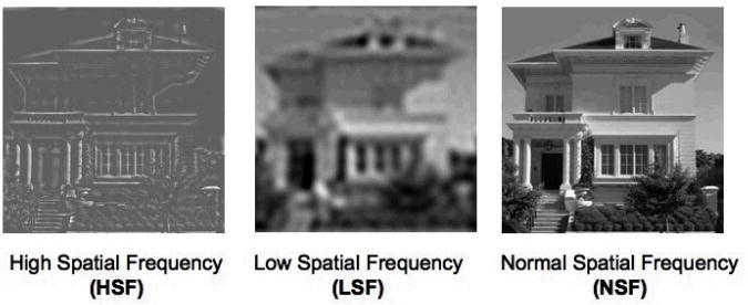
Method: Fourteen medication-free participants with DSM-IV BDD and 14 healthy controls participated. We performed functional magnetic resonance imaging (fMRI) while participants matched photographs of houses that were unaltered, contained only high spatial frequency (HSF, high detail) information or only low spatial frequency (LSF, low detail) information. The primary outcome was group differences in blood oxygen level-dependent (BOLD) signal changes.
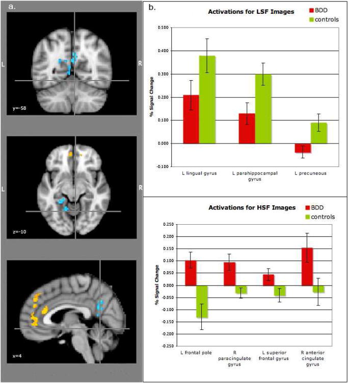
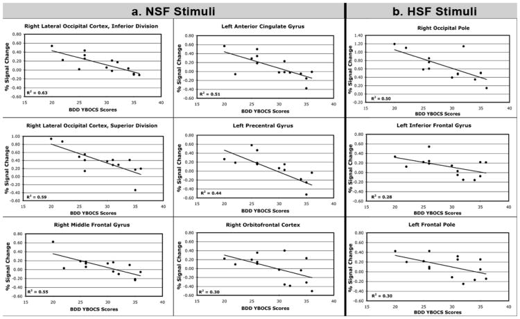
Results: The BDD group showed lower activity in the parahippocampal gyrus, lingual gyrus and precuneus for LSF images. There were greater activations in medial prefrontal regions for HSF images, although no significant differences when compared to a low-level baseline. Greater symptom severity was associated with lower activity in the dorsal occipital cortex and ventrolateral prefrontal cortex for normal spatial frequency (NSF) and HSF images.
Conclusions: Individuals with BDD have abnormal brain activation patterns when viewing objects. Hypoactivity in visual association areas for configural and holistic (low detail) elements and abnormal allocation of prefrontal systems for details are consistent with a model of imbalances in global versus local processing. This may occur not only for appearance but also for general stimuli unrelated to their symptoms.
Conflict of interest statement
The authors have no conflicts of interest to report.
Figures
Similar articles
-
Abnormalities of visual processing and frontostriatal systems in body dysmorphic disorder.Arch Gen Psychiatry. 2010 Feb;67(2):197-205. doi: 10.1001/archgenpsychiatry.2009.190. Arch Gen Psychiatry. 2010. PMID: 20124119 Free PMC article.
-
Visual information processing of faces in body dysmorphic disorder.Arch Gen Psychiatry. 2007 Dec;64(12):1417-25. doi: 10.1001/archpsyc.64.12.1417. Arch Gen Psychiatry. 2007. PMID: 18056550
-
Anorexia nervosa and body dysmorphic disorder are associated with abnormalities in processing visual information.Psychol Med. 2015 Jul;45(10):2111-22. doi: 10.1017/S0033291715000045. Epub 2015 Feb 5. Psychol Med. 2015. PMID: 25652023 Free PMC article.
-
Visual processing in anorexia nervosa and body dysmorphic disorder: similarities, differences, and future research directions.J Psychiatr Res. 2013 Oct;47(10):1483-91. doi: 10.1016/j.jpsychires.2013.06.003. Epub 2013 Jun 27. J Psychiatr Res. 2013. PMID: 23810196 Free PMC article. Review.
-
A hierarchy of visual processing deficits in body dysmorphic disorder: a conceptual review and empirical investigation.Cogn Neuropsychiatry. 2024 Mar;29(2):116-140. doi: 10.1080/13546805.2024.2326243. Epub 2024 Apr 2. Cogn Neuropsychiatry. 2024. PMID: 38563811 Review.
Cited by
-
Study protocol of comprehensive risk evaluation for anorexia nervosa in twins (CREAT): a study of discordant monozygotic twins with anorexia nervosa.BMC Psychiatry. 2020 Oct 14;20(1):507. doi: 10.1186/s12888-020-02903-7. BMC Psychiatry. 2020. PMID: 33054774 Free PMC article.
-
Effects of visual attention modulation on dynamic functional connectivity during own-face viewing in body dysmorphic disorder.Neuropsychopharmacology. 2021 Oct;46(11):2030-2038. doi: 10.1038/s41386-021-01039-w. Epub 2021 May 28. Neuropsychopharmacology. 2021. PMID: 34050267 Free PMC article.
-
Neural Correlates of Own- and Other-Face Perception in Body Dysmorphic Disorder.Front Psychiatry. 2020 Apr 24;11:302. doi: 10.3389/fpsyt.2020.00302. eCollection 2020. Front Psychiatry. 2020. PMID: 32395110 Free PMC article.
-
Body dysmorphic disorder.Nat Rev Dis Primers. 2024 Dec 5;10(1):92. doi: 10.1038/s41572-024-00577-z. Nat Rev Dis Primers. 2024. PMID: 39639018 Free PMC article. Review.
-
Comparison of visual perceptual organization in schizophrenia and body dysmorphic disorder.Psychiatry Res. 2015 Sep 30;229(1-2):426-33. doi: 10.1016/j.psychres.2015.05.107. Epub 2015 Jun 27. Psychiatry Res. 2015. PMID: 26184989 Free PMC article.
References
-
- Aguirre GK, Zarahn E, D'Esposito M. The variability of human, BOLD hemodynamic responses. Neuroimage. 1998;8:360–9. - PubMed
-
- American Psychiatric Association. Diagnostic and statistical manual of mental disorders : DSM-IV-TR. American Psychiatric Association; Washington, DC: 2000.
-
- Beckmann M, Jenkinson M, Smith S. General multi-level linear modeling for group analysis in FMRI. Neuroimage. 2003;20:1052–1063. - PubMed
-
- Bird G, Catmur C, Silani G, Frith C, Frith U. Attention does not modulate neural responses to social stimuli in autism spectrum disorders. Neuroimage. 2006;31:1614–24. - PubMed
Publication types
MeSH terms
Grants and funding
LinkOut - more resources
Full Text Sources
