Review
. 2011 Apr;21(2):228-37.
doi: 10.1016/j.conb.2011.02.007.

Wiring and rewiring of the retinogeniculate synapse

Affiliations
Review

Wiring and rewiring of the retinogeniculate synapse

Y Kate Hong et al. Curr Opin Neurobiol. 2011 Apr.

Abstract

The formation and refinement of synaptic circuits are areas of research that have fascinated neurobiologists for decades. A recurrent theme seen at many CNS synapses is that neuronal connections are at first imprecise, but refine and can be rearranged with time or with experience. Today, with the advent of new technologies to map and monitor neuronal circuits, it is worthwhile to revisit a powerful experimental model for examining the development and plasticity of synaptic circuits--the retinogeniculate synapse.


Figures

Figure 1
Figure 1. Developmental phases of retinogeniculate connectivity in mouse
In the retina (red lines), spontaneous activity in the form of cholinergic waves is followed by glutamatergic waves [5]. Eye-opening in mice occurs around postnatal day 12–14 (arrowhead). Visually evoked activity begins shortly before this, when light can be first detected through closed eyelids and persists throughout adulthood. Development of retinal connections to the dorsal lateral geniculate (LGN) can be divided into 3 phases. During phase I (black), axon refinement occurs during retinopic refinement and eye-specific segregation. Throughout the second phase (blue), synaptic connections are further refined through continued elimination and strengthening of synapses. The third phase (green) involves the stabilization and maintenance of established connections. This period encompasses a period in which retinogeniculate connectivity can be influenced by visual experience.
Figure 2
Figure 2. Laminar patterns of axon terminal projections in the mouse LGN
A. The location of the dorsal LGN in a mouse coronal section (green). B. Schematic representation of axon terminal zones in the LGN of transgenic mouse lines that selectively label functional subsets of RGCs including: JAMB, OFF-direction selective RGCs; CB2, transient OFF-α RGCs; DRD4-RGCs, ONOFF direction selective RGCs; and BD, ON-OFF direction selective RGCs [–14]. Colored regions represent regions of the LGN that contained labeled axons of the different RGC subsets in the mature LGN. It is still unclear how these territories change over development. Identical contours of a coronal LGN section were drawn for simplicity and may represent different rostrocaudal sections.
Figure 3
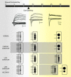
Figure 3
Functional development of the mouse retinogeniculate synapse. (Top) Representative excitatory synaptic responses to incremental increases in optic nerve stimulation from P11, 17 and 28 mice [23]. Synaptic currents are recorded while alternating the holding potential between −70 mV (inward currents) and +40 mV (outward currents) to assess both AMPAR and NMDAR currents, respectively. (Bottom) Schematic representation of synaptic refinement is drawn to symbolize the experimental data for number of inputs and synaptic strength at different points in development [26,30]. Under normal conditions, synaptic strength increases by 20-fold and afferent inputs decrease from more than 10 down to just 1–3. An RGC input is symbolized by a horizontal line connected to a black circle; the size of the circle represents the relative strength of the input. Each black circle represents an axon making numerous contacts (release sites) with the postsynaptic neuron. Sensory deprivation by dark rearing animals from birth (chronic dark rear) shows no significant difference to normally reared mice. However, dark rearing after a period of visual experience (late dark rear), results in an increase in number of RGC inputs and weakening of established connections. During this period, these changes can be reversed when the animal is re-exposed to light (late dark rear + recovery).
Figure 4
Figure 4
Structural development of retinal axon terminals in the LGN of the cat. We revisit these classic single-RGC morphology studies to compare the anatomical changes seen over development with the changes in retinogeniculate synaptic strength and innervation observed from mice. Similar single axon studies in mice are currently not available, thus comparisons are made relative to the time of completion of eye specific segregation and eye opening. (Top) Schematic representation of the progression of eye-specific segregation in the cat. Projections from one eye colored in red, the other in green (see top right box). Yellow region represents region of overlap between the two eyes (Bottom) Illustration of morphological changes of single RGC axon arbors described from classic studies in the cat. Bottom right box shows the location of the arbor within the LGN, Images of arbors adapted from [20] (embryonic (E) time points) and [62] (adult).

Similar articles

Cited by

References

    1. Lu T, Trussell LO. Development and elimination of endbulb synapses in the chick cochlear nucleus. J Neurosci. 2007;27:808–817. - PMC - PubMed
    1. Wang H, Zhang ZW. A critical window for experience-dependent plasticity at whisker sensory relay synapse in the thalamus. J Neurosci. 2008;28:13621–13628. - PMC - PubMed
    1. Kano M, Hashimoto K. Synapse elimination in the central nervous system. Curr Opin Neurobiol. 2009;19:154–161. - PubMed
    1. Pfeiffenberger C, Yamada J, Feldheim DA. Ephrin-As and patterned retinal activity act together in the development of topographic maps in the primary visual system. J Neurosci. 2006;26:12873–12884. - PMC - PubMed
    1. Huberman AD, Feller MB, Chapman B. Mechanisms underlying development of visual maps and receptive fields. Annu Rev Neurosci. 2008;31:479–509. - PMC - PubMed

Publication types