. 2011 Jul 8;286(27):23708-16.
doi: 10.1074/jbc.M111.241265. Epub 2011 May 10.

Studies of the genetics, function, and kinetic mechanism of TagE, the wall teichoic acid glycosyltransferase in Bacillus subtilis 168

Affiliations

Studies of the genetics, function, and kinetic mechanism of TagE, the wall teichoic acid glycosyltransferase in Bacillus subtilis 168

Sarah E Allison et al. J Biol Chem. .

Abstract

The biosynthetic enzymes involved in wall teichoic acid biogenesis in gram-positive bacteria have been the subject of renewed investigation in recent years with the benefit of modern tools of biochemistry and genetics. Nevertheless, there have been only limited investigations into the enzymes that glycosylate wall teichoic acid. Decades-old experiments in the model gram-positive bacterium, Bacillus subtilis 168, using phage-resistant mutants implicated tagE (also called gtaA and rodD) as the gene coding for the wall teichoic acid glycosyltransferase. This study and others have provided only indirect evidence to support a role for TagE in wall teichoic acid glycosylation. In this work, we showed that deletion of tagE resulted in the loss of α-glucose at the C-2 position of glycerol in the poly(glycerol phosphate) polymer backbone. We also reported the first kinetic characterization of pure, recombinant wall teichoic acid glycosyltransferase using clean synthetic substrates. We investigated the substrate specificity of TagE using a wide variety of acceptor substrates and found that the enzyme had a strong kinetic preference for the transfer of glucose from UDP-glucose to glycerol phosphate in polymeric form. Further, we showed that the enzyme recognized its polymeric (and repetitive) substrate with a sequential kinetic mechanism. This work provides direct evidence that TagE is the wall teichoic acid glycosyltransferase in B. subtilis 168 and provides a strong basis for further studies of the mechanism of wall teichoic acid glycosylation, a largely uncharted aspect of wall teichoic acid biogenesis.


Figures

FIGURE 1.
FIGURE 1.
Proposed pathway for wall teichoic acid biosynthesis in B. subtilis 168. Wall teichoic acid polymers are composed of a disaccharide-containing N-acetylglucosamine 1-phosphate (white oval with phosphate group) and N-acetylmannosamine (black oval) and ∼40 repeating glycerol 3-phosphate units (square). These polymers are synthesized on the cytoplasmic face of the cell membrane on an undecaprenyl phosphate molecule (wavy line with phosphate group). Once synthesis is complete, the polymer is exported to the outside of the cell and covalently attached to peptidoglycan.
FIGURE 2.
FIGURE 2.
Deletion of tagE leads to the loss of α-glucose at the C-2 position of poly(glycerol phosphate) wall teichoic acid. Shown are phosphate analysis (A) and SPO1 phage susceptibility (B) of the ΔtagE (EB2252) and the wild-type B. subtilis 168 parent strain (EB6). C, 1H NMR spectra of wall teichoic acid isolated from B. subtilis 168 (top) and the ΔtagE strain (bottom). The α-glucose anomeric resonance at δ5.07 is indicated by a dashed vertical line. Error bars, S.D.
SCHEME 1.
SCHEME 1.
Reaction catalyzed by TagE in vivo. In the in vitro TagE activity assay, the lipid β portion of the poly(glycerol phosphate) polymer has been replaced by a soluble analog of lipid β (N-acetylmannosamine-β-(1–4)-GlcNAc-1-phosphate-phosphate-tridecane).
FIGURE 3.
FIGURE 3.
Dependence of TagE activity on time and enzyme concentration. Reactions contained 3 mm UDP-glucose, 15 μm lipid ϕ.40 analog, and 1 (●), 2.5 (○), 5 (▴), or 10 nm (△) TagE. Reactions were quenched with urea to a final concentration of 6 m following 1-, 3-, 6-, and 12-min incubations. The conversion of UDP-glucose to UDP was monitored at 262 nm following separation by paired ion HPLC. Inset, plot of initial velocity versus TagE concentration. The slope of the plot represents the turnover of TagE under saturating conditions (16 s−1). Error bars, S.D.
FIGURE 4.
FIGURE 4.
TagE utilizes a sequential (ternary complex) mechanism. A, double reciprocal plot of 1/velocity versus 1/[UDP-glucose]. UDP-glucose was varied from 1600 to 12,800 μm while the lipid ϕ.40 concentration was fixed at 0.5 (●), 1 (○), 2 (■), and 8 μm (□). B, double reciprocal plot of initial rate data with varying lipid ϕ.40 analog concentrations (0.5–8 μm) at fixed UDP-glucose concentrations (1600 (●), 3200 (○), 6400 (■), and 12,800 μm (□)). All experiments were conducted with 2.5 nm TagE, and reaction rates were determined by monitoring the conversion of UDP-glucose to UDP at 262 nm. The data were fitted by the non-linear least squares method to a sequential kinetic mechanism (Equation 2).
FIGURE 5.
FIGURE 5.
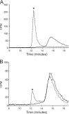
Glycosylation impairs poly(glycerol phosphate) polymerization by TagF in vitro. A, the solid black trace indicates elution for the 14C-lipid ϕ.5 analog. The dotted black trace indicates 14C-lipid ϕ.5 analog elution for a reaction containing CDP-glycerol and TagF. Formation of a higher molecular weight product, whose elution is consistent with a polymer containing nearly 50 glycerol phosphate units, is indicated by an asterisk. B, elution of the glycosylated 14C-lipid ϕ.5 analog following incubation with (dotted line) or without (solid line) TagF. Glycosylated and non-glycosylated 14C-lipid ϕ.5 analogues were incubated with 4 mm CDP-glycerol and 100 nm TagF for 5 h. Reaction substrates and products were then separated by size exclusion chromatography on a Waters Shodex KW-803 column in buffer containing 0.1% ammonium hydrogen carbonate and 10% acetonitrile at 0.5 ml/min.
FIGURE 6.
FIGURE 6.
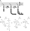
Mechanism of wall teichoic acid glycosylation in B. subtilis 168 in vivo. A, TagF synthesizes a polymer of ∼40 units of glycerol 3-phosphate (square) from CDP-glycerol onto lipid ϕ.1. Once polymer synthesis is complete or nearly complete TagE transfers glucose (light gray oval) from UDP-glucose onto the poly(glycerol phosphate) polymer. The extent and distribution of glucose along the polymer are unknown. The modified polymer is then exported by the ABC transporter, TagGH. B, a proposed SN1-like reaction for TagE involves formation of an oxocarbenium ion intermediate and nucleophilic attack from the acceptor substrate on the same face from which the leaving group departs. The products of the reaction are a glycosylated poly(glycerol phosphate) polymer and UDP. R, an oxygen or NH2 group on an amino acid in the active site of the enzyme; R1, the poly(glycerol phosphate) acceptor substrate.

References

    1. D'Elia M. A., Millar K. E., Beveridge T. J., Brown E. D. (2006) J. Bacteriol. 188, 8313–8316 - PMC - PubMed
    1. Weidenmaier C., Kokai-Kun J. F., Kristian S. A., Chanturiya T., Kalbacher H., Gross M., Nicholson G., Neumeister B., Mond J. J., Peschel A. (2004) Nat. Med. 10, 243–245 - PubMed
    1. D'Elia M. A., Henderson J. A., Beveridge T. J., Heinrichs D. E., Brown E. D. (2009) J. Bacteriol. 191, 4030–4034 - PMC - PubMed
    1. Neuhaus F. C., Baddiley J. (2003) Microbiol. Mol. Biol. Rev. 67, 686–723 - PMC - PubMed
    1. Peschel A., Otto M., Jack R. W., Kalbacher H., Jung G., Götz F. (1999) J. Biol. Chem. 274, 8405–8410 - PubMed

Publication types

MeSH terms

LinkOut - more resources