Clinical Trial
. 2011 Oct;31(10):2086-95.
doi: 10.1038/jcbfm.2011.72. Epub 2011 May 11.

Bilateral transcranial direct current stimulation modulates activation-induced regional blood flow changes during voluntary movement

Affiliations
Clinical Trial

Bilateral transcranial direct current stimulation modulates activation-induced regional blood flow changes during voluntary movement

Caroline Paquette et al. J Cereb Blood Flow Metab. 2011 Oct.

Abstract

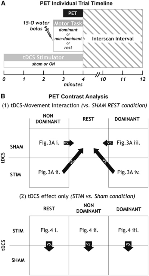
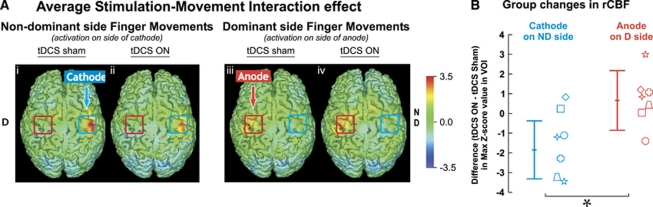
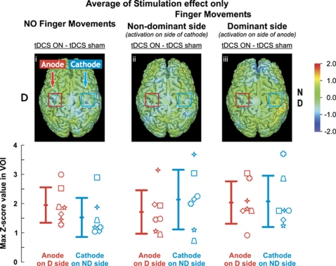
Transcranial direct current stimulation (tDCS) is a noninvasive brain stimulation technique that induces changes in cortical excitability: anodal stimulation increases while cathodal stimulation reduces excitability. Imaging studies performed after unilateral stimulation have shown conflicting results regarding the effects of tDCS on surrogate markers of neuronal activity. The aim of this study was to directly measure these effects on activation-induced changes in regional cerebral blood flow (ΔrCBF) using positron emission tomography (PET) during bilateral tDCS. Nine healthy subjects underwent repeated rCBF measurements with (15)O-water and PET during a simple motor task while receiving tDCS or sham stimulation over the primary motor cortex (M1). Motor evoked potentials (MEPs) were also assessed before and after real and sham stimulation. During tDCS with active movement, ΔrCBF in M1 was significantly lower on the cathodal than the anodal side when compared with sham stimulation. This decrease in ΔrCBF was accompanied by a decrease in MEP amplitude on the cathodal side. No effect was observed on resting or activated rCBF relative to sham stimulation. We thus conclude that it is the interaction of cathodal tDCS with activation-induced ΔrCBF rather than the effect on resting or activated rCBF itself which constitutes the physiological imaging correlate of tDCS.


Figures

Figure 1
Figure 1
Illustration of the measurement used for transcranial direct current stimulation (tDCS) electrode placement over the first dorsal interosseous muscle (FDI) hotspot, as determined by functional magnetic resonance imaging (fMRI). The antero-posterior distance of the M1 representation from the nasion was first measured after the subject's skull on the midline. The lateral component of the electrodes was then measured from the midline on each side. These measures were used to position the tDCS electrodes on the subject.
Figure 2
Figure 2
Positron emission tomography (PET) experimental set-up. Individual trial timeline is shown in (A). The contrasts tested are shown in (B) for (1) transcranial direct current stimulation (tDCS)–movement interaction, contrasting conditions with tDCS SHAM during rest condition and (2) for stimulation effects only, where tDCS STIM was contrasted with tDCS SHAM for each of the three motor tasks. Each contrast is shown as Z-transformed difference images averaged across subjects in Figures 3 and 4, as indicated in the respective boxes. STIM, stimulation.
Figure 3
Figure 3
Regional cerebral blood flow (rCBF) changes with bilateral transcranial direct current stimulation (tDCS) during finger movements. Z-transformed difference images averaged across seven subjects are shown in (A). Activations are shown during finger movements on the nondominant (i and ii) and dominant (iii and iv) hand. Differences are obtained from contrast with the sham rest condition. The red (anode) and blue (cathode) squares drawn on each image correspond to the tDCS electrodes. (B) Mean (±1s.d.) group changes in rCBF during cathodal and anodal stimulation are shown. Individual subjects are represented by the seven different symbols. *P<0.05, one-tailed paired t-test.
Figure 4
Figure 4
Regional cerebral blood flow (rCBF) changes with bilateral transcranial direct current stimulation (tDCS) only. Z-transformed differences images averaged across seven subjects are shown above and group means (±1s.d.) are shown in the bottom row. Activations are shown during rest (versus sham rest) on the left panel (i), during movements of the nondominant hand (versus sham nondominant movement) in center panel (ii) and for the dominant hand (versus sham dominant movement) in the right panel (iii). No change was observed in ΔrCBF when contrasting tDCS stimulation versus sham with nondominant (center panel) or dominant (right panel) finger movements. The red (anode) and blue (cathode) squares drawn on each image correspond to the tDCS electrodes. Individual subjects are represented by the seven different symbols.
Figure 5
Figure 5
Changes in motor evoked potentials (MEPs) after bilateral transcranial direct current stimulation (tDCS). Values for sham on the cathodal and anodal side were pooled together.

References

    1. Ardolino G, Bossi B, Barbieri S, Priori A. Non-synaptic mechanisms underlie the after-effects of cathodal transcutaneous direct current stimulation of the human brain. J Physiol. 2005;568:653–663. - PMC - PubMed
    1. Baudewig J, Nitsche MA, Paulus W, Frahm J. Regional modulation of BOLD MRI responses to human sensorimotor activation by transcranial direct current stimulation. Magn Reson Med. 2001;45:196–201. - PubMed
    1. Benninger DH, Lomarev M, Lopez G, Wassermann EM, Li X, Considine E, Hallett M. Transcranial direct current stimulation for the treatment of Parkinson's disease. J Neurol Neurosurg Psychiatry. 2010;81:1105–1111. - PMC - PubMed
    1. Bindman LJ, Lippold OC, Redfearn JW. The action of brief polarizing currents on the cerebral cortex of the rat (1) during current flow and (2) in the production of long-lasting after-effects. J Physiol. 1964;172:369–382. - PMC - PubMed
    1. Boggio PS, Castro LO, Savagim EA, Braite R, Cruz VC, Rocha RR, Rigonatti SP, Silva MT, Fregni F. Enhancement of non-dominant hand motor function by anodal transcranial direct current stimulation. Neurosci Lett. 2006;404:232–236. - PubMed

Publication types

LinkOut - more resources