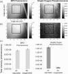
Dual-channel imaging system for singlet oxygen and photosensitizer for PDT
- PMID: 21559134
- PMCID: PMC3087579
- DOI: 10.1364/BOE.2.001233
Dual-channel imaging system for singlet oxygen and photosensitizer for PDT
Abstract
A two-channel optical system has been developed to provide spatially resolved simultaneous imaging of singlet molecular oxygen ((1)O(2)) phosphorescence and photosensitizer (PS) fluorescence produced by the photodynamic process. The current imaging system uses a spectral discrimination method to differentiate the weak (1)O(2) phosphorescence that peaks near 1.27 μm from PS fluorescence that also occurs in this spectral region. The detection limit of (1)O(2) emission was determined at a concentration of 500 nM benzoporphyrin derivative monoacid (BPD) in tissue-like phantoms, and these signals observed were proportional to the PS fluorescence. Preliminary in vivo images with tumor laden mice indicate that it is possible to obtain simultaneous images of (1)O(2) and PS tissue distribution.
Keywords: (170.0110) Imaging systems; (170.3880) Medical and biological imaging; (170.5180) Photodynamic therapy.
Figures


References
-
- Weishaupt K. R., Gomer C. J., Dougherty T. J., “Identification of singlet oxygen as the cytotoxic agent in photoinactivation of a murine tumor,” Cancer Res. 36(7 PT 1), 2326–2329 (1976). - PubMed
-
- Kubler A. C., “Photodynamic Therapy,” Med. Laser Appl. 20(1), 37–45 (2005).10.1016/j.mla.2005.02.001 - DOI
-
- S. L. Jacques, “Simple theory, measurements, and rules of thumb for dosimetry during photodynamic therapy,” Proc. SPIE 1065, 100–108 (1989).
-
- B. W. Pogue, R. D. Braun, J. L. Lanzen, C. Erikson, and M. W. Dewhirst, “Oxygen microelectrode measurements in R3230Ac Tumors during photodynamic therapy with verteporfin,” Proc. SPIE 4248, 144 (2001).
Grants and funding
LinkOut - more resources
Full Text Sources
Other Literature Sources
