. 2011 Apr 29;6(4):e19305.
doi: 10.1371/journal.pone.0019305.

Modeling initiation of Ewing sarcoma in human neural crest cells

Affiliations

Modeling initiation of Ewing sarcoma in human neural crest cells

Cornelia von Levetzow et al. PLoS One. .

Abstract

Ewing sarcoma family tumors (ESFT) are aggressive bone and soft tissue tumors that express EWS-ETS fusion genes as driver mutations. Although the histogenesis of ESFT is controversial, mesenchymal (MSC) and/or neural crest (NCSC) stem cells have been implicated as cells of origin. For the current study we evaluated the consequences of EWS-FLI1 expression in human embryonic stem cell-derived NCSC (hNCSC). Ectopic expression of EWS-FLI1 in undifferentiated hNCSC and their neuro-mesenchymal stem cell (hNC-MSC) progeny was readily tolerated and led to altered expression of both well established as well as novel EWS-FLI1 target genes. Importantly, whole genome expression profiling studies revealed that the molecular signature of established ESFT is more similar to hNCSC than any other normal tissue, including MSC, indicating that maintenance or reactivation of the NCSC program is a feature of ESFT pathogenesis. Consistent with this hypothesis, EWS-FLI1 induced hNCSC genes as well as the polycomb proteins BMI-1 and EZH2 in hNC-MSC. In addition, up-regulation of BMI-1 was associated with avoidance of cellular senescence and reversible silencing of p16. Together these studies confirm that, unlike terminally differentiated cells but consistent with bone marrow-derived MSC, NCSC tolerate expression of EWS-FLI1 and ectopic expression of the oncogene initiates transition to an ESFT-like state. In addition, to our knowledge this is the first demonstration that EWS-FLI1-mediated induction of BMI-1 and epigenetic silencing of p16 might be critical early initiating events in ESFT tumorigenesis.


Conflict of interest statement

Competing Interests: The authors have declared that no competing interests exist.

Figures

Figure 1
Figure 1. hNCSC have mesenchymal differentiation potential.
(A) Within 2 days of transfer to adherent plates p75+ hNCSC initiated a change from small, NCSC-like to larger MSC-like cells. By 5 days all cells were mesenchymal in appearance (In all images scale bars = 100 µm). (B) Oil-Red O- and Alizarin Red staining after 3 weeks in defined media confirmed that hNCSC can be induced to differentiate into adipocytes and osteoblasts, respectively, albeit less robustly than BM-MSC. Simliarly stained BM-MSC that had been exposed to standard growth media are shown as negative controls. (C) RT-PCR showed that hNCSC express the mesenchymal marker VIM (vimentin) as well as NCSC genes B3GAT1 (HNK-1), NGFR (p75), and NES (nestin). In contrast, BM-MSC (3 different lines as detailed in methods) do not express NCSC genes. BM-MSC: bone marrow-derived MSC; hNCSC: hESC-derived NCSC.
Figure 2
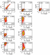
Figure 2. Flow cytometry confirms MSC markers in adherent hNCSC.
hNCSC were isolated from in vitro differentiating hESC using p75-FACS. Nearly 50% of the cells also expressed the MSC marker CD73 on Day 0. After 2 days in adherent conditions the morphology of isolated cells changed from small cuboidal cells to larger mesenchymal cells (see Fig. 1A). Consistent with this morphologic change flow cytometric analysis shows that after 5 days in culture most cells continue to express p75 but, in addition, nearly all cells express the MSC-associated markers CD73, CD105, CD90, CD29, and CD44.
Figure 3
Figure 3. hNCSC and hNC-MSC tolerate expression of EWS-FLI1.
(A) pCLS-EWS-FLI1-V5-2A-EGFP lentiviral expression vector. (B) hNCSC transduced with either control (CV)- or EWS-FLI1-vectors (EF) formed neurospheres in non-adherent culture (phase contrast (top) and GFP-filters (bottom) 100×). (C) RT-PCR confirmed expression of EWS-FLI1 in EF cells and also induction of known EWS-FLI1 target genes NKX2.2 and ID2. (Results are shown for 3 independent experiments). (D) Cell cycle was assessed in adherent EGFP+ hNC-MSC 6 weeks after lentiviral transduction. Western blot (left) confirmed continued robust expression of the EWS-FLI1-V5 protein and flow cytometric analysis of DNA content (right) showed continued proliferation and minimal cell death in both EF and CV cells in 2 independent experiments.
Figure 4
Figure 4. Whole genome expression profiling confirms that ESFT cell lines and tumors are most closely related to hNCSC.
(A) Unsupervised 3D principal components analysis (PCA) of freshly isolated hNCSC (N = 3), hNC-MSC (N = 3), adult bone marrow-derived MSC (BM-MSC; N = 3), 10 primary ESFT, 10 ESFT cell lines and 11 adult tissues (each in triplicate) shows that ESFT cluster more closely with stem cells than any adult tissue and are most similar to undifferentiated hNCSC. Notably testes and cerebellar tissues cluster separately from other adult tissues (breast, heart, kidney, liver, muscle, pancreas, prostate, spleen, thyroid). All 17,881 core transcripts represented on the array were included for this analysis. (B) Unsupervised clustering of samples in (A) was repeated after exclusion of 1,676 EWS-FLI1 target genes. ESFT continue to cluster with hNCSC.
Figure 5
Figure 5. EWS-FLI1 induces a NCSC phenotype in hNC-MSC.
(A) EWS-FLI1 (EF-NC) and control vector (CV-NC) transduced hNC-MSC were passaged in serum-containing media for 3 weeks and morphology imaged by immunocytochemistry. EF-NC cells were smaller and less mesenchymal in appearance than CV-NC cells (scale bar = 100 µm). (B) RT-PCR confirms down regulation of NCSC genes B3GAT1 (HNK-1) and NGFR (p75) in CV-NC cells after 2 weeks. In contrast, NCSC and EWS-FLI1 target gene expression remained high in EF-NC cells (results for 3 independent experiments are shown). (C) Transduced, EGFP+ cells were isolated 6 weeks after transduction and analyzed by western blot. Data from 3 independent experiments show consistent up regulation of BMI-1 and EZH2 and repression of p16 in EF-NC cells. (D) After 6 weeks BMI-1, EZH2 and p16 expression in EF-NC cells were equivalent to freshly isolated hNCSC. In contrast, CV-NC cells down regulated polycomb proteins and up regulated p16.
Figure 6
Figure 6. EWS-FLI1 transduced human neural crest cells avoid cellular senescence.
(A) Senescence-associated beta-galactosidase staining 11 weeks post-transduction shows that CV-NC but not EF-NC cells had undergone senescence (scale bar = 100 µm) (B) Functional p53 was confirmed in EF-NC cells by induction of p53 and p21 proteins following gamma irradiation. (C) Senescence-resistant EF-NC retain the ability to form neurospheres (scale bar = 100 µm).
Figure 7
Figure 7. Inhibition of BMI-1 in EF-NC cells restores p16 expression and leads to cellular senescence.
(A) Inducible BMI-1 knockdown lentiviral vector. (B) Red fluorescent protein expression following doxycyline treatment confirmed induction of inert non-silencing (NS) and BMI-1-targeted (BMI1 kd) shRNA in transduced cells (scale bar = 100 µm). (C) Western blot analysis confirmed BMI-1 knockdown in BMI1 kd cells accompanied by de-repression of p16 and concomitant down regulation of EZH2. (D) BMI-1 knockdown cells also expressed reduced levels of Cyclin D1 compared to control (NS) cells. (E) Senescence-associated beta-galactosidase staining 1 week post-doxycyline induction shows that BMI-1 kd cells were senescent (scale bar = 100 µm).

References

    1. Riggi N, Stamenkovic I. The Biology of Ewing sarcoma. Cancer Lett. 2007;254:1–10. - PubMed
    1. Kovar H. Downstream EWS/FLI1 - upstream Ewing's sarcoma. Genome Med. 2010;2:8. - PMC - PubMed
    1. Deneen B, Denny CT. Loss of p16 pathways stabilizes EWS/FLI1 expression and complements EWS/FLI1 mediated transformation. Oncogene. 2001;20:6731–6741. - PubMed
    1. Lessnick SL, Dacwag CS, Golub TR. The Ewing's sarcoma oncoprotein EWS/FLI induces a p53-dependent growth arrest in primary human fibroblasts. Cancer Cell. 2002;1:393–401. - PubMed
    1. Huang HY, Illei PB, Zhao Z, Mazumdar M, Huvos AG, et al. Ewing sarcomas with p53 mutation or p16/p14ARF homozygous deletion: a highly lethal subset associated with poor chemoresponse. J Clin Oncol. 2005;23:548–558. - PubMed

Publication types