

Model sensitivity and use of the comparative finite element method in mammalian jaw mechanics: mandible performance in the gray wolf
- PMID: 21559475
- PMCID: PMC3084775
- DOI: 10.1371/journal.pone.0019171
Model sensitivity and use of the comparative finite element method in mammalian jaw mechanics: mandible performance in the gray wolf
Abstract
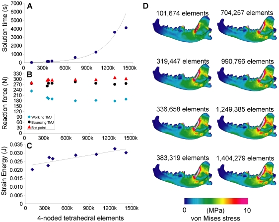
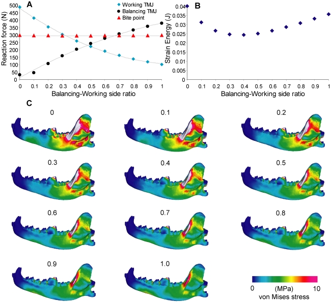
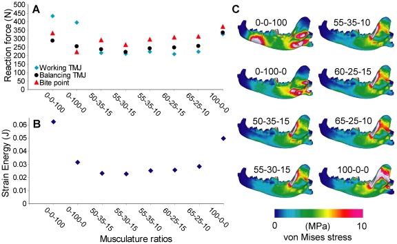
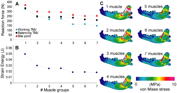
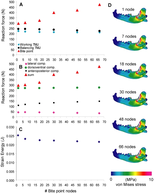
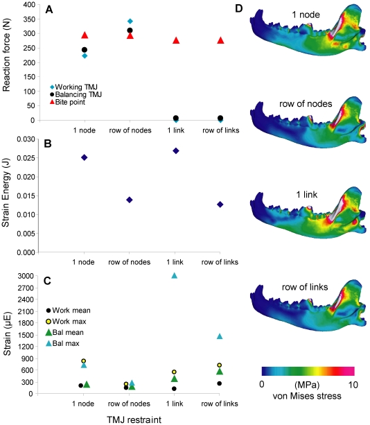
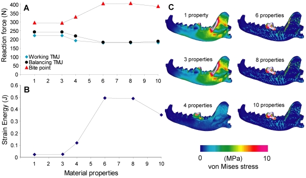
Finite Element Analysis (FEA) is a powerful tool gaining use in studies of biological form and function. This method is particularly conducive to studies of extinct and fossilized organisms, as models can be assigned properties that approximate living tissues. In disciplines where model validation is difficult or impossible, the choice of model parameters and their effects on the results become increasingly important, especially in comparing outputs to infer function. To evaluate the extent to which performance measures are affected by initial model input, we tested the sensitivity of bite force, strain energy, and stress to changes in seven parameters that are required in testing craniodental function with FEA. Simulations were performed on FE models of a Gray Wolf (Canis lupus) mandible. Results showed that unilateral bite force outputs are least affected by the relative ratios of the balancing and working muscles, but only ratios above 0.5 provided balancing-working side joint reaction force relationships that are consistent with experimental data. The constraints modeled at the bite point had the greatest effect on bite force output, but the most appropriate constraint may depend on the study question. Strain energy is least affected by variation in bite point constraint, but larger variations in strain energy values are observed in models with different number of tetrahedral elements, masticatory muscle ratios and muscle subgroups present, and number of material properties. These findings indicate that performance measures are differentially affected by variation in initial model parameters. In the absence of validated input values, FE models can nevertheless provide robust comparisons if these parameters are standardized within a given study to minimize variation that arise during the model-building process. Sensitivity tests incorporated into the study design not only aid in the interpretation of simulation results, but can also provide additional insights on form and function.
Conflict of interest statement
Figures
References
-
- Rayfield EJ. Finite element analysis and understanding the biomechanics and evolution of living and fossil organisms. Annual Review of Earth and Planetary Science. 2007;35:541–576.
-
- Ross CF. Finite element analysis in vertebrate biomechanics. The Anatomical Record. 2005;283A:253–258. - PubMed
-
- Kupczik K. Virtual biomechanics: basic concepts and technical aspects of finite element analysis in vertebrate morphology. Journal of Anthropological Sciences. 2008;86:193–198. - PubMed
-
- Fastnacht M, Hess N, Frey E, Weiser H-P. Finite element analysis in vertebrate palaeontology. Senckenbergiana lethaea. 2002;82:195–206.
-
- McHenry C, Clausen PD, Daniel WJT, Meers MB, Pendharkar A. Biomechanics of the rostrum in crocodilians, a comparative analysis using finite element modeling. The Anatomical Record. 2006;288A:827–849. - PubMed
Publication types
MeSH terms
LinkOut - more resources
Full Text Sources
Other Literature Sources
