

A first- and second-order motion energy analysis of peripheral motion illusions leads to further evidence of "feature blur" in peripheral vision
- PMID: 21559513
- PMCID: PMC3084698
- DOI: 10.1371/journal.pone.0018719
A first- and second-order motion energy analysis of peripheral motion illusions leads to further evidence of "feature blur" in peripheral vision
Abstract
Background: Anatomical and physiological differences between the central and peripheral visual systems are well documented. Recent findings have suggested that vision in the periphery is not just a scaled version of foveal vision, but rather is relatively poor at representing spatial and temporal phase and other visual features. Shapiro, Lu, Huang, Knight, and Ennis (2010) have recently examined a motion stimulus (the "curveball illusion") in which the shift from foveal to peripheral viewing results in a dramatic spatial/temporal discontinuity. Here, we apply a similar analysis to a range of other spatial/temporal configurations that create perceptual conflict between foveal and peripheral vision.
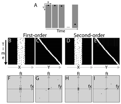
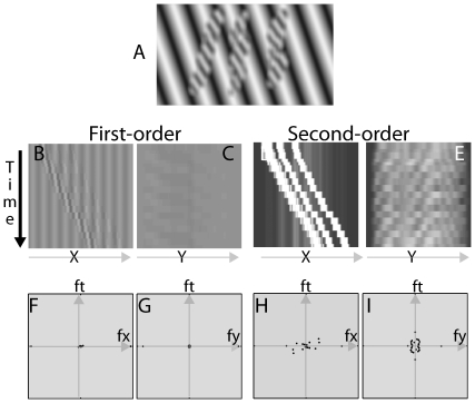
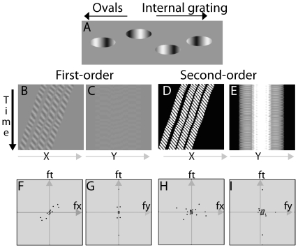
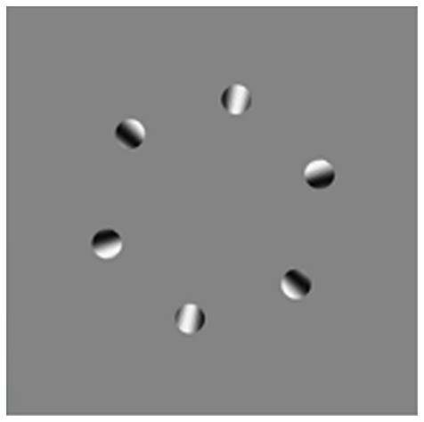
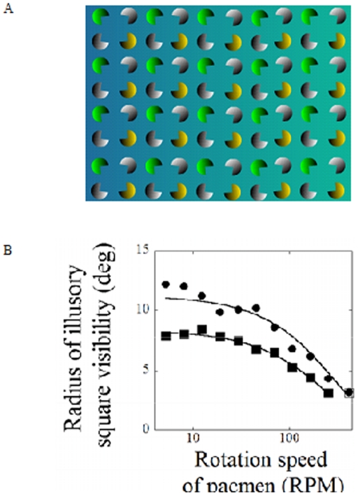
Methodology/principal findings: To elucidate how the differences between foveal and peripheral vision affect super-threshold vision, we created a series of complex visual displays that contain opposing sources of motion information. The displays (referred to as the peripheral escalator illusion, peripheral acceleration and deceleration illusions, rotating reversals illusion, and disappearing squares illusion) create dramatically different perceptions when viewed foveally versus peripherally. We compute the first-order and second-order directional motion energy available in the displays using a three-dimensional Fourier analysis in the (x, y, t) space. The peripheral escalator, acceleration and deceleration illusions and rotating reversals illusion all show a similar trend: in the fovea, the first-order motion energy and second-order motion energy can be perceptually separated from each other; in the periphery, the perception seems to correspond to a combination of the multiple sources of motion information. The disappearing squares illusion shows that the ability to assemble the features of Kanisza squares becomes slower in the periphery.
Conclusions/significance: The results lead us to hypothesize "feature blur" in the periphery (i.e., the peripheral visual system combines features that the foveal visual system can separate). Feature blur is of general importance because humans are frequently bringing the information in the periphery to the fovea and vice versa.
Conflict of interest statement
Figures

Similar articles
-
Transitions between central and peripheral vision create spatial/temporal distortions: a hypothesis concerning the perceived break of the curveball.PLoS One. 2010 Oct 13;5(10):e13296. doi: 10.1371/journal.pone.0013296. PLoS One. 2010. PMID: 20967247 Free PMC article.
-
The furrow illusion: peripheral motion becomes aligned with stationary contours.J Vis. 2012 Nov 20;12(12):12. doi: 10.1167/12.12.12. J Vis. 2012. PMID: 23169994
-
The Uniformity Illusion.Psychol Sci. 2017 Jan;28(1):56-68. doi: 10.1177/0956797616672270. Epub 2016 Nov 15. Psychol Sci. 2017. PMID: 28078975
-
A review of interactions between peripheral and foveal vision.J Vis. 2020 Nov 2;20(12):2. doi: 10.1167/jov.20.12.2. J Vis. 2020. PMID: 33141171 Free PMC article. Review.
-
Motion perception in the peripheral visual field.Perception. 1982;11(4):457-62. doi: 10.1068/p110457. Perception. 1982. PMID: 6763677 Review.
Cited by
-
The Role of Feature Tracking in the Furrow Illusion.Front Hum Neurosci. 2016 Mar 7;10:81. doi: 10.3389/fnhum.2016.00081. eCollection 2016. Front Hum Neurosci. 2016. PMID: 27014018 Free PMC article.
-
Following Randolph Blake's furrow further.J Vis. 2025 May 1;25(6):9. doi: 10.1167/jov.25.6.9. J Vis. 2025. PMID: 40408119 Free PMC article.
-
Robust natural depth for anticorrelated random dot stereogram for edge stimuli, but minimal reversed depth for embedded circular stimuli, irrespective of eccentricity.PLoS One. 2022 Sep 22;17(9):e0274566. doi: 10.1371/journal.pone.0274566. eCollection 2022. PLoS One. 2022. PMID: 36137132 Free PMC article.
-
Dissociation of neuronal and psychophysical responses to local and global motion.Curr Biol. 2011 Dec 6;21(23):2023-8. doi: 10.1016/j.cub.2011.10.049. Epub 2011 Dec 5. Curr Biol. 2011. PMID: 22153156 Free PMC article.
-
What you see is what you get: motor resonance in peripheral vision.Exp Brain Res. 2015 Oct;233(10):3013-22. doi: 10.1007/s00221-015-4371-0. Epub 2015 Jul 14. Exp Brain Res. 2015. PMID: 26169105
References
-
- Curcio CA, Sloan KR, Kalina RE, Henderickson AE. Human photoreceptor topography. J Comp Neurology. 1990;292:497–523. - PubMed
-
- Wässle H, Grünert U, Röhrenbeck J, Boycott BB. Cortical magnification factor and the ganglion cell density of the primate retina. Nature. 1989;341:643–646. - PubMed
-
- Masland RH. The fundamental plan of the retina. Nature Neuroscience. 2001;4:877–886. - PubMed
-
- Martin PR, Grünert U. Ganglion Cells in Mammalian Retinae. In: Chalupa LM, Werner JS, editors. The Visual Neurosciences. Cambridge: MIT Press; 2004.
-
- Tootell RB, Silverman MS, Switkes E, De Valois RL. Deoxyglucose analysis of retinotopic organization in primate striate cortex. Science. 1982;218:902–904. - PubMed
Publication types
MeSH terms
Grants and funding
LinkOut - more resources
Full Text Sources
Miscellaneous
