

Porcine model of early onset scoliosis based on animal growth created with posterior mini-invasive spinal offset tethering: a preliminary report
- PMID: 21559770
- PMCID: PMC3207335
- DOI: 10.1007/s00586-011-1830-6
Porcine model of early onset scoliosis based on animal growth created with posterior mini-invasive spinal offset tethering: a preliminary report
Abstract
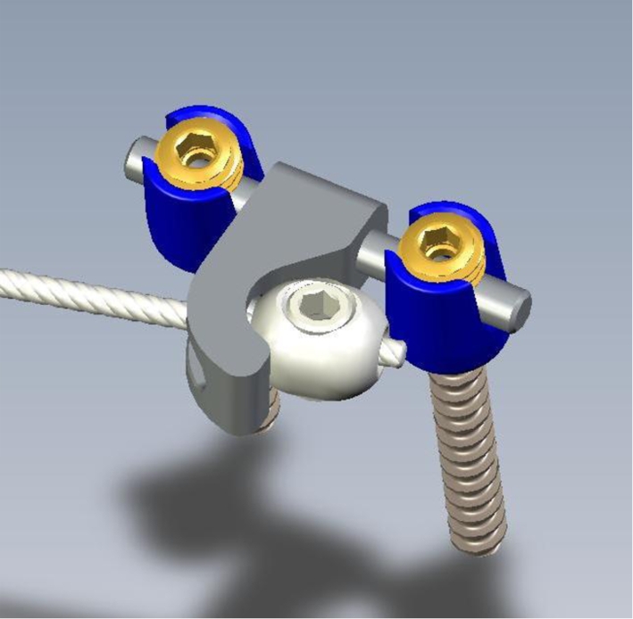
Several models of scoliosis were developed in the past 10 years. In most of them, deformations are induced in old animals and required long time observation period and a chest wall ligation ± resection. The purpose of the study was to create a scoliosis model with a size similar to an early onset scoliosis and an important growth potential without chest wall injuring. An original offset implant was fixed posteriorly and connected with a cable in seven (6 + 1 control) one-month-old Landrace pigs. The mean initial spinal length (T1-S1) was 25 cm and the mean weight was 9 kg. After 2 months observation, spinal deformities were assessed with a three dimension stereographic analysis. In four animals, the cable was sectioned and the deformities followed-up for next 2 months. No post-operative complication was observed. Mean weight growth was 10 kg/month and mean spine lengthening (T1-S1) was 7 cm/month. In 2 months, we obtained structural scoliotic curves with vertebral and disk wedging which were maximal at the apex of the curve. Mean frontal and sagittal Cobb angles was 45°. Chest wall associated deformities were similar to those observed in scoliotic deformities and were correlated to spinal deformities (p = 0.03). The cable section resulted in a partial curve regression influenced by disk elasticity and could probably be influenced by gravity loads (Decrease of the Cobb angle of 30% in the sagittal plane and 45% in the frontal plane). According to the results, the model creates a structural scoliosis and chest wall deformity that is similar to an early onset scoliosis. The spinal deformities were obtained quickly, and were consistent between animals in term of amount and characteristic.
Figures


Similar articles
-
A porcine early-onset scoliosis model created using a posterior mini-invasive method: a pilot study.J Spinal Disord Tech. 2014 Dec;27(8):E294-300. doi: 10.1097/BSD.0000000000000117. J Spinal Disord Tech. 2014. PMID: 25374380
-
[Correlation study between spinal curvatures and vertebral and disk deformities in idiopathic scoliosis].Ann Chir. 1999;53(8):798-807. Ann Chir. 1999. PMID: 10584392 French.
-
Posterior vertebral column resection for correction of rigid spinal deformity curves greater than 100°.J Neurosurg Spine. 2012 Dec;17(6):540-51. doi: 10.3171/2012.9.SPINE111026. Epub 2012 Oct 12. J Neurosurg Spine. 2012. PMID: 23062175
-
The course of sagittal plane abnormality in the patients with congenital scoliosis managed with convex growth arrest.Spine (Phila Pa 1976). 2004 Mar 1;29(5):547-52; discussion 552-3. doi: 10.1097/01.brs.0000106493.54636.b4. Spine (Phila Pa 1976). 2004. PMID: 15129069 Review.
-
A comparison of growth among growth-friendly systems for scoliosis: a systematic review.Spine J. 2019 May;19(5):789-799. doi: 10.1016/j.spinee.2018.08.017. Epub 2018 Oct 2. Spine J. 2019. PMID: 30290228
Cited by
-
Animal models for scoliosis research: state of the art, current concepts and future perspective applications.Eur Spine J. 2013 Mar;22 Suppl 2(Suppl 2):S81-95. doi: 10.1007/s00586-012-2396-7. Epub 2012 Oct 26. Eur Spine J. 2013. PMID: 23099524 Free PMC article. Review.
-
Biomechanical analysis of the camelid cervical intervertebral disc.J Orthop Translat. 2014 Dec 23;3(1):34-43. doi: 10.1016/j.jot.2014.12.001. eCollection 2015 Jan. J Orthop Translat. 2014. PMID: 30035038 Free PMC article.
-
Pressure distributions inside intervertebral discs under unilateral pedicle screw fixation in a porcine spine model.J Orthop Surg Res. 2018 Oct 16;13(1):254. doi: 10.1186/s13018-018-0962-3. J Orthop Surg Res. 2018. PMID: 30326934 Free PMC article.
-
Safe corridor for the implantation of thoracolumbar pedicle screws in growing pigs: A morphometric study.PLoS One. 2017 Oct 23;12(10):e0184857. doi: 10.1371/journal.pone.0184857. eCollection 2017. PLoS One. 2017. PMID: 29059193 Free PMC article.
-
Pedicle Screw Fixation Study in Immature Porcine Spines to Improve Pullout Resistance during Animal Testing.PLoS One. 2015 Oct 9;10(10):e0127463. doi: 10.1371/journal.pone.0127463. eCollection 2015. PLoS One. 2015. PMID: 26451947 Free PMC article.
References
-
- Dickson R. Early-onset idiopathic scoliosis. In: Weinstein S, editor. The pediatric spine: principles and practice. New York: Raven Press; 1994.
-
- Roach J. Adolescent idiopathic scoliosis: non surgical treatment. In: Weinstein S, editor. The pediatric spine: principles and practice. New York: Raven Press; 1994.
Publication types
MeSH terms
LinkOut - more resources
Full Text Sources
Medical
Miscellaneous
