. 2012 Feb;132(1):109-19.
doi: 10.1007/s10549-011-1568-1. Epub 2011 May 11.

ErbB1/2 tyrosine kinase inhibitor mediates oxidative stress-induced apoptosis in inflammatory breast cancer cells

Affiliations

ErbB1/2 tyrosine kinase inhibitor mediates oxidative stress-induced apoptosis in inflammatory breast cancer cells

Katherine M Aird et al. Breast Cancer Res Treat. 2012 Feb.

Abstract

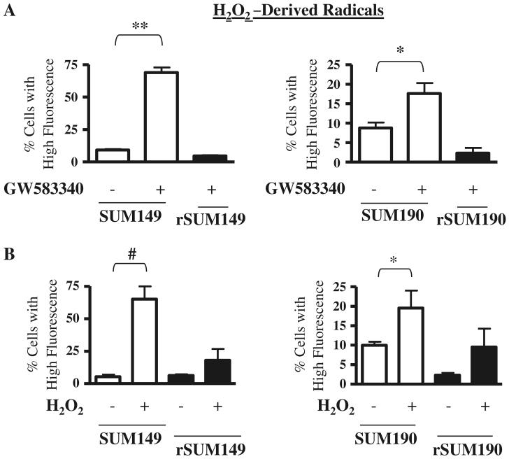
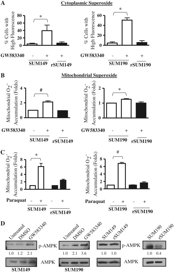
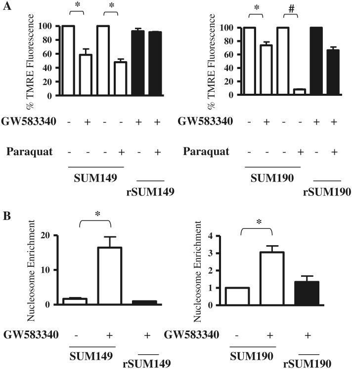
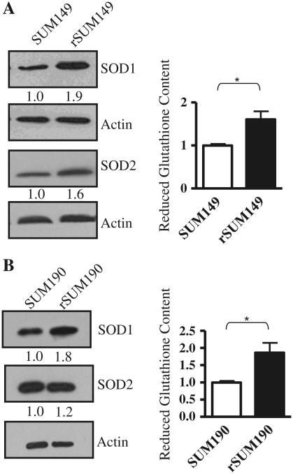
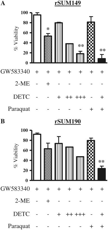
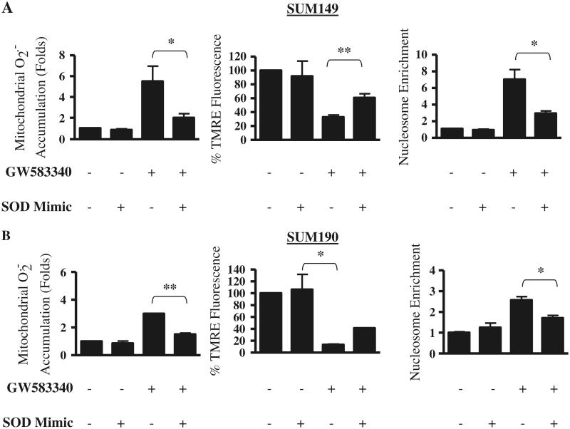
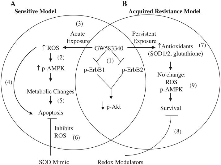
Overexpression of epidermal growth factor receptors (ErbB) is frequently seen in inflammatory breast cancer (IBC). Treatment with ErbB1/2-targeting agents (lapatinib) mediates tumor apoptosis by downregulating ErbB1/2 phosphorylation and downstream survival signaling. In this study, using carboxy-H(2)DCFDA, DHE, and MitoSOX Red to examine changes in hydrogen peroxide radicals, cytoplasmic and mitochondrial superoxide, respectively, we observed that GW583340 (a lapatinib-analog) increases reactive oxygen species (ROS) in two models of IBC (SUM149, SUM190) that are sensitive to ErbB1/2 blockade. This significant increase in ROS levels was similar to those generated by classical oxidative agents H(2)O(2) and paraquat. In contrast, minimal to basal levels of ROS were measured in a clonal population of GW583340-resistant IBC cells (rSUM149 and rSUM190). The GW583340-resistant IBC cells displayed increased SOD1, SOD2, and glutathione expression, which correlated with decreased sensitivity to the apoptotic-inducing effects of GW583340, H(2)O(2), and paraquat. The ROS increase and cell death in the GW583340-sensitive cells was reversed by simultaneous treatment with a superoxide dismutase (SOD) mimic. Additionally, overcoming the high levels of antioxidants using redox modulators induced apoptosis in the GW583340-resistant cells. Taken together, these data demonstrate a novel mechanism of lapatinib-analog-induced apoptosis and indicate that resistant cells have increased antioxidant potential, which can be overcome by treatment with SOD modulators.


Figures

Fig. 1
Fig. 1
Effect of GW583340 and H2O2 on H2O2-derived radical accumulation in IBC cells a SUM149 (left panel) and SUM190 (right panel) cells were treated with 7.5 and 2.5 μM GW583340, respectively, for 24 h, and H2O2-derived radical accumulation was compared to rSUM149 and rSUM190 cells growing in GW583340. b SUM149 and rSUM149 cells were treated with 500 μM H2O2 (left panel) and SUM190 and rSUM190 cells were treated with 100 μM H2O2 (right panel), and H2O2-derived radical accumulation was assessed. Bars represent mean ± SEM of the percentage of cells with high carboxy-H2DCFDA fluorescence (n = 3; *P < 0.05, **P < 0.01, #P < 0.005)
Fig. 2
Fig. 2
Effect of GW583340 and paraquat on superoxide accumulation in IBC cells a SUM149 (left panel) and SUM190 (right panel) cells were treated with 7.5 and 2.5 μM GW583340, respectively, for 24 h, and cytoplasmic superoxide accumulation was compared to rSUM149 and rSUM190 cells growing in GW583340. Bars represent mean ± SEM of the percentage of cells with high DHE fluorescence (n = 3; *P < 0.05). b SUM149 (left panel) and SUM190 (right panel) cells were treated with 7.5 and 2.5 μM GW583340, respectively, for 24 h, and mitochondrial superoxide accumulation was compared to rSUM149 and rSUM190 cells growing in GW583340. Bars represent mean ± SEM MitoSOX Red fluorescence normalized to DMSO (n = 3; *P < 0.05, #P < 0.005). c SUM149 and rSUM149 cells were treated with 5 mM paraquat (left panel) and SUM190 and rSUM190 cells were treated with 0.5 mM paraquat (right panel), and superoxide accumulation was assessed. Bars represent mean ± SEM MitoSOX Red fluorescence normalized to DMSO (n = 3; *P < 0.05, #P < 0.005). d p-AMPK expression in SUM149 and SUM190 cells treated for 1 h with 7.5 and 2.5 μM GW583340, respectively (left panel). p-AMPK expression was assessed in rSUM149 and rSUM190 cells growing in GW583340 (right panel) and compared to their parental counterparts. Blots were stripped and reprobed for total AMPK. Numbers represent densitometric analysis of p-AMPK normalized to total AMPK levels
Fig. 3
Fig. 3
Effect of GW583340 and paraquat on mitochondrial membrane potential and apoptosis of IBC cells. Parental cells treated with GW583340 (SUM149: 7.5 μM GW583340; SUM190: 2.5 μM GW583340) were compared to rSUM149 and rSUM190 cells growing in the same concentration of GW583340. Mitochondrial membrane potential (a) and apoptosis (b) were assessed by TMRE staining and nucleosome enrichment, respectively (n = 2, *P < 0.05, #P < 0.005)
Fig. 4
Fig. 4
Antioxidant expression in parental and GW583340-resistant IBC cells a Left panel immunoblot analysis of SOD1 and SOD2 in rSUM149 vs. SUM149 cells. Actin was used as a loading control. Numbers represent densitometric analysis of SOD1 or SOD2 normalized to actin. Right panel reduced glutathione content in SUM149 and rSUM149 cells. Bars represent mean ± SEM luciferase RLU normalized to SUM149 (n = 2; *P < 0.05). b Left panel immunoblot analysis of SOD1 and SOD2 in rSUM190 vs. SUM190 cells. Actin was used as a loading control. Numbers represent densitometric analysis of SOD1 or SOD2 normalized to actin. Right panel reduced glutathione content in SUM190 and rSUM190 cells. Bars represent mean ± SEM luciferase RLU normalized to SUM190 (n = 2; *P < 0.05)
Fig. 5
Fig. 5
Effect of redox modulators on viability of rSUM149 and rSUM190 IBC cells. rSUM149 (a) and rSUM190 (b) cells growing in GW583340 (white bar) were treated with 1 μM 2-ME (dotted bar), 10–100 μM DETC (striped bars: +, 10 μM; ++, 50 μM; +++, 100 μM), paraquat (checked bar), and paraquat and 10 μM DETC in combination (black bars), and viability was assessed using trypan blue exclusion. Bars represent mean ± SEM% viable cells normalized to control cells (n = 2; *P < 0.05, **P < 0.01)
Fig. 6
Fig. 6
Effect of GW583340 and SOD mimic alone and in combination on ROS production, mitochondrial membrane potential, and apoptosis in parental IBC cells. SUM149 (a) and SUM190 (b) cells were treated with 30 μM SOD mimic and 20 μM GW583340 alone or in combination for 24 h, and mitochondrial superoxide generation (left panels), mitochondrial membrane potential (middle panels), and apoptosis (right panels) were assessed using MitoSOX Red, TMRE, and cytoplasmic nucleosome enrichment, respectively. Bars represent mean ± SEM normalized to untreated or DMSO (n = 2; *P < 0.05, **P < 0.005)
Fig. 7
Fig. 7
Schematic representation of mechanism of action of GW583340 in sensitive (a) and acquired resistance (b) IBC models. (1) GW583340 binds to and inhibits phosphorylation of ErbB1 and ErbB2 and subsequently decreases p-AKT expression in both sensitive and acquired resistance models [4, 11, 12]. However, in sensitive cells, GW583340 treatment causes an acute stress by increasing ROS (3), which in turn can increase AMPK activation (2) [20]. Both a substantial increase in ROS [42] and activation of AMPK [8, 16] and changes in metabolism [8] can independently lead to apoptosis of sensitive cells (4, 5). Treatment of sensitive cells in the presence of a SOD mimic can reverse this ROS-induced apoptosis (6). In contrast, cells with acquired GW583340 resistance have higher antioxidant expression (7) corresponding with no change in ROS accumulation or p-AMPK activation (9) in the presence of GW583340 and cell survival. Modulation of the redox pathway using 2-ME or DETC can sensitize these cells to ROS-generating agents(8)

References

    1. Anderson WF, Schairer C, Chen BE, Hance KW, Levine PH. Epidemiology of inflammatory breast cancer (IBC) Breast Dis. 2005;22:9–23. - PMC - PubMed
    1. Woodward WA, Cristofanilli M. Inflammatory breast cancer. Semin Radiat Oncol. 2009;19:256–265. - PubMed
    1. Van den Eynden GG, Van der Auwera I, Van Laere S, Colpaert CG, van Dam P, Merajver S, Kleer CG, Harris AL, Van Marck EA, Dirix LY, et al. Validation of a tissue microarray to study differential protein expression in inflammatory and non-inflammatory breast cancer. Breast Cancer Res Treat. 2004;85:13–22. - PubMed
    1. Xia W, Mullin RJ, Keith BR, Liu LH, Ma H, Rusnak DW, Owens G, Alligood KJ, Spector NL. Anti-tumor activity of GW572016: a dual tyrosine kinase inhibitor blocks EGF activation of EGFR/erbB2 and downstream Erk1/2 and AKT pathways. Oncogene. 2002;21:6255–6263. - PubMed
    1. Xia W, Bisi J, Strum J, Liu L, Carrick K, Graham KM, Treece AL, Hardwicke MA, Dush M, Liao Q, et al. Regulation of survivin by ErbB2 signaling: therapeutic implications for ErbB2-overexpressing breast cancers. Cancer Res. 2006;66:1640–1647. - PubMed

Publication types

MeSH terms