Review
. 2011 Jul;68(13):2215-29.
doi: 10.1007/s00018-011-0714-6. Epub 2011 May 11.

Paneth cell α-defensins in enteric innate immunity

Affiliations
Review

Paneth cell α-defensins in enteric innate immunity

André Joseph Ouellette. Cell Mol Life Sci. 2011 Jul.

Abstract

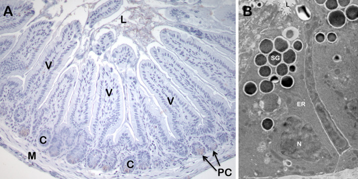
Paneth cells at the base of small intestinal crypts of Lieberkühn secrete high levels of α-defensins in response to cholinergic and microbial stimuli. Paneth cell α-defensins are broad spectrum microbicides that function in the extracellular environment of the intestinal lumen, and they are responsible for the majority of secreted bactericidal peptide activity. Paneth cell α-defensins confer immunity to oral infection by Salmonella enterica serovar Typhimurium, and they are major determinants of the composition of the small intestinal microbiome. In addition to host defense molecules such as α-defensins, lysozyme, and Pla2g2a, Paneth cells also produce and release proinflammatory mediators as components of secretory granules. Disruption of Paneth cell homeostasis, with subsequent induction of endoplasmic reticulum stress, autophagy, or apoptosis, contributes to inflammation in diverse genetic and experimental mouse models.


Figures

Fig. 1
Fig. 1
The small intestinal Paneth cell. a A section of ileum from an AKR strain mouse was lightly stained with hematoxylin–eosin. The position of Paneth cells (PC) at the base of crypts (C) is indicated by arrows. Also shown are the small intestinal lumen (L), representative villi (V), and small intestinal smooth muscle (M). b Large, electron-dense secretory granules (SG) of a Paneth cell. The SG are clustered at the apical cell membrane in proximity of the crypt lumen (L). The extensive endoplasmic reticulum (ER) and basolaterally-oriented nucleus (N), characteristic of this lineage are also indicated. Dr. Susan J. Hagen, Beth Israel Deaconess Medical Center, Boston, MA, provided the electron micrograph
Fig. 2
Fig. 2
Selected constituents of Paneth cell dense core secretory granules. Arrows denote electron dense secretory granules (G) near the apical surface of a mouse Paneth cell. The dense core granules, surrounded by an electron lucent matrix, are released apically into the lumen, labeled as such, of the crypt by uncharacterized vesicular fusion events. In contrast to the highly fucosylated N- and O-linked glycoprotein components of the electron dense cores, the electron lucent peripheral halo contains GalNAc residues in O-linked oligosaccharides and terminal GlcNAc residues in N- and O-linked glycoconjugates [178]. Box at right contains a partial listing of biologically active proteins confirmed to be constituents of Paneth cell granules
Fig. 3
Fig. 3
Structures of α-, β- and θ-defensins. The backbone and disulfide structures are shown of RK-1 (upper left), a monomeric rabbit α-defensin, HNP-3 (upper right), a dimeric human α-defensin, hBD-1 (lower left), a monomeric human β-defensin, and RTD-1 (lower right), a θ-defensin from rhesus macaque [179]. Reprinted from [180], with permission
Fig. 4
Fig. 4
Primary structures of representative α-defensins. The single letter notation amino acid sequences of human Paneth cell α-defensins HD-5 and HD-6, human neutrophil α-defensins HNPs 1 and 4, mouse Paneth cell α-defensins cryptdins 1–6, rhesus macaque myeloid α-defensins RMADs 1–8, and rhesus macaque Paneth cell α-defensins REDs 1–6 are aligned. The canonical 1–6, 2–4, 3–5 pairings of the conserved Cys residue positions in all sequences are noted above the HD5 primary structure, and conserved Arg, Glu, and Gly residues are denoted within boxes. Dash characters were introduced into the HD6 and Crp4 sequences to maintain the alignment of conserved residue positions
Fig. 5
Fig. 5
The characteristic α-defensin fold. a Diagram of the backbone traces of the 20 lowest energy NMR structures of the mouse Paneth cell α-defensin cryptdin-4 (Crp4). b Ribbon diagram of the Crp4 structure showing the triple β-sheet in turquoise and the three disulfide bonds shown in gold, as generated with MOLMOL. c, d Overlay of the NMR structure of Crp4 (blue) with the NMR structure of rabbit kidney RK-1 (c, red) and the crystal structure of human HNP-3 (d, red). Reprinted from [59], with permission
Fig. 6
Fig. 6
Alignment of CRS1C-1, CRS4C-1, and Crp1 precursors. a Schematic representation of the structure of Paneth cell α-defensin genes and precursors. Reprinted with permission from [, Fig. 1]. b The primary structures deduced Crp1, CRS4C-1, and CRS1C-1 precursors are aligned, beginning at the initiating methionine residue position, with the signal sequence shown in yellow text and the proregion in aqua text. Natural proCrp1 and CRS4C have been isolated and their MMP7 processed N-termini have been confirmed experimentally [78, 144]. In the lower half of the figure, Cys residues in the mature peptides (orange text) are underlined and highlighted in white text to denote the differences between the Crp1 α-defensin Cys distribution and that of the CRS1C and CRS4C peptide subfamilies
Fig. 7
Fig. 7
Phylogenetic relationships between rat and mouse α-defensin, CRS1C, and CRS4C genes. The introns of mouse Paneth cell α-defensin, CRS1C and CRS4C genes from the NIH C57BL/6 and the mixed strain Celera assemblies, introns of rat enteric α-defensin genes, and second introns of rat myeloid α-defensin genes, and vestigial mouse myeloid α-defensin (DefmaN-ps) genes were used to construct the phylogenetic tree. The tree was rooted with the intron of the human α-defensin-5 (HD-5) gene, and construction of the tree involved the calculation of the proportion difference (p-distance) of aligned nucleotide sites of the entire intron sequences according to the neighbor-joining method. One thousand bootstrap replications were used to test the reliability of each branch. Solid lines maintain phylogenetic distances, but dashed lines do not in order to maintain legibility of sequences of the tree. Reprinted from [90], with permission

References

    1. Porter EM, Bevins CL, Ghosh D, Ganz T. The multifaceted Paneth cell. Cell Mol Life Sci. 2002;59:156–170. doi: 10.1007/s00018-002-8412-z. - DOI - PMC - PubMed
    1. Cheng H, Merzel J, Leblond CP. Renewal of Paneth cells in the small intestine of the mouse. Am J Anat. 1969;126:507–525. doi: 10.1002/aja.1001260409. - DOI - PubMed
    1. Cheng H. Origin, differentiation and renewal of the four main epithelial cell types in the mouse small intestine. IV. Paneth cells. Am J Anat. 1974;141:521–535. doi: 10.1002/aja.1001410406. - DOI - PubMed
    1. Cheng H, Leblond CP. Origin, differentiation and renewal of the four main epithelial cell types in the mouse small intestine. V. Unitarian Theory of the origin of the four epithelial cell types. Am J Anat. 1974;141:537–561. doi: 10.1002/aja.1001410407. - DOI - PubMed
    1. Cheng H, Leblond CP. Origin, differentiation and renewal of the four main epithelial cell types in the mouse small intestine. I. Columnar cell. Am J Anat. 1974;141:461–479. doi: 10.1002/aja.1001410403. - DOI - PubMed

Publication types

Substances

LinkOut - more resources