. 2011 Dec;24(10):1302-12.
doi: 10.1002/nbm.1692. Epub 2011 May 11.

A brain MRI study of chronic fatigue syndrome: evidence of brainstem dysfunction and altered homeostasis

Affiliations

A brain MRI study of chronic fatigue syndrome: evidence of brainstem dysfunction and altered homeostasis

Leighton R Barnden et al. NMR Biomed. 2011 Dec.

Abstract

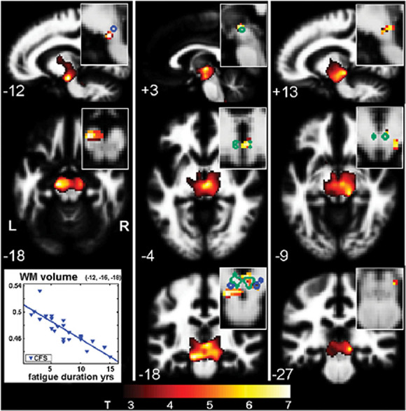
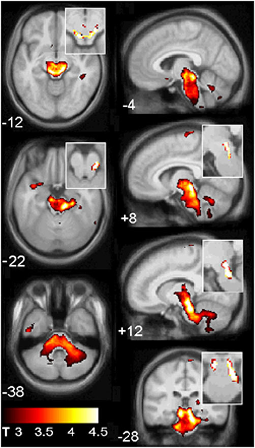
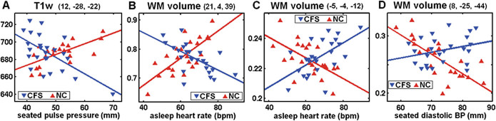
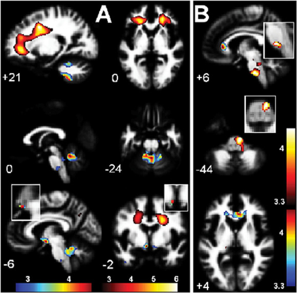
To explore brain involvement in chronic fatigue syndrome (CFS), the statistical parametric mapping of brain MR images has been extended to voxel-based regressions against clinical scores. Using SPM5 we performed voxel-based morphometry (VBM) and analysed T(1) - and T(2) -weighted spin-echo MR signal levels in 25 CFS subjects and 25 normal controls (NC). Clinical scores included CFS fatigue duration, a score based on the 10 most common CFS symptoms, the Bell score, the hospital anxiety and depression scale (HADS) anxiety and depression, and hemodynamic parameters from 24-h blood pressure monitoring. We also performed group × hemodynamic score interaction regressions to detect locations where MR regressions were opposite for CFS and NC, thereby indicating abnormality in the CFS group. In the midbrain, white matter volume was observed to decrease with increasing fatigue duration. For T(1) -weighted MR and white matter volume, group × hemodynamic score interactions were detected in the brainstem [strongest in midbrain grey matter (GM)], deep prefrontal white matter (WM), the caudal basal pons and hypothalamus. A strong correlation in CFS between brainstem GM volume and pulse pressure suggested impaired cerebrovascular autoregulation. It can be argued that at least some of these changes could arise from astrocyte dysfunction. These results are consistent with an insult to the midbrain at fatigue onset that affects multiple feedback control loops to suppress cerebral motor and cognitive activity and disrupt local CNS homeostasis, including resetting of some elements of the autonomic nervous system (ANS).


Figures

Figure 1
Figure 1
Total age-adjusted absolute grey matter volumes in the brainstem region of interest (ROI) versus seated pulse pressure (PP). The regression line is shown for (CFS) (p = 0.0008). The dashed red line indicates the normal control (NC) mean. There was no significant regression for NC.
Figure 2
Figure 2
Voxel-based morphometry (VBM) white matter (WM) volume versus fatigue duration (Table 3, item 3). The cluster was formed with uncorrected voxel p < 0.005. Sections are through the three peak voxels with corrected voxel p < 0.05 and are located in the corticospinal tract (left column), red nucleus/hypothalamus periventricular area (centre) and medial geniculate body (right). Magnified inserts improve localization of the peaks via a voxel threshold of uvP < 0.00001 and, for anatomic guidance, include edges from an atlas of the substantia nigra (blue) and red nucleus (green). The plot shows relative WM volume versus fatigue duration at the peak voxel and indicates shrinkage of 1% per year. The background image is the mean WM image from the present study.
Figure 3
Figure 3
T1w results from a group × seated pulse pressure (PP) interaction (Table 3, item 1) involving the midbrain and hindbrain, shown on the mean gradient echo sections from the present study. The cluster was formed with a threshold of uvP = 0.005. Magnified inserts with a more stringent threshold (uvP < 0.0001) locate the peak significance to the midbrain reticular substance and periaqueductal grey matter.
Figure 4
Figure 4
Plots, for group × haemodynamic score interactions, of values for each chronic fatigue syndrome (CFS) and normal control (NC) subject at the most significant voxel in the four clusters described in Table 3, items 1, 2, 8, 5. Individual MR values have been adjusted for age and global value. The lines are the general linear model fits. The mean MR values in the CFS and NC groups are not significantly different. The voxels were located: (A) in the low midbrain reticular substance (Fig. 3); (B) in prefrontal white matter (WM) (Fig. 5A); (C) in the hypothalamus (Fig. 5A); and (D) in the caudal basal pons (Fig. 5A). Although only one of these individual voxel interactions (B) was significant after correction for multiple comparisons, all four interactions extended across clusters that were significant.
Figure 5
Figure 5
White matter (WM) volume interaction results for (A). Group × asleep heart rate (Table 3, items 2, 7, 8 and Fig. 4B,C), and (B). Group × seated diastolic blood pressure (BP) (Table 3, item 5) interaction regressions. In (A) significant clusters formed with a uvP = 0.005 threshold were observed bilaterally in deep prefrontal WM and in the cerebellar vermis and hypothalamus. In (B) a significant cluster formed with a uvP = 0.005 threshold is seen in the caudal basal pons. The two colour bars distinguish between different polarity interactions (e.g. Fig. 4B, C). Thus, in (A) red–yellow–white indicates where as asleep heart rate (HR) increases, WM volume decreases in chronic fatigue syndrome (CFS) but increases in normal controls (NC), whereas blue–green–red indicates the reverse. Magnified inserts locate the clusters in the hypothalamus (A) and pons (B) with a more stringent threshold of p = 0.0005. The background image is the mean white matter image from the present study.

References

    1. Fukuda K, Straus SE, Hickie I, Sharpe MC, Dobbins JG, Komaroff A. The chronic fatigue syndrome: a comprehensive approach to its definition and study. Ann. Intern. Med. 1994;121:953–959. - PubMed
    1. Michiels V, Cluydts R. Neuropsychological functioning in chronic fatigue syndrome: a review. Acta Psychiatr. Scand. 2001;103:84–93. - PubMed
    1. de Lange F, Kalkman J, Bleijenberg G, Hagoort P, Sieberen P, van der Werf S, et al. Neural correlates of the chronic fatigue syndrome - an fMRI study. Brain. 2004;127:1948–1957. - PubMed
    1. Newton J, Okonkwo O, Sutcliffe K, Seth A, Shin J, Jones D. Symptoms of autonomic dysfunction in chronic fatigue syndrome. Q. J. Med. 2007;100:519–526. - PubMed
    1. Natelson B, Weaver S, Tseng C-L, Ottenweller J. Spinal fluid abnormalities in patients with chronic fatigue syndrome. Clin. Diag. Lab. Immun. 2005;12:52–55. - PMC - PubMed

Publication types