

Case Reports
doi: 10.3109/17453674.2011.581265.
Epub 2011 May 11.
Biofilms in chronic diabetic foot ulcers--a study of 2 cases
Affiliations
- PMID: 21561305
- PMCID: PMC3235322
- DOI: 10.3109/17453674.2011.581265
Item in Clipboard
Case Reports
Biofilms in chronic diabetic foot ulcers--a study of 2 cases
Acta Orthop.
2011 Jun.
No abstract available
Figures
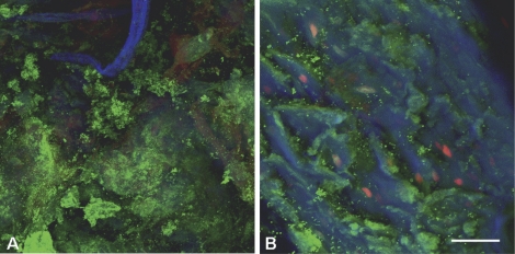
CLSM images of biofilm on soft tissue from patient 1. Overlay projection (includes all the slices in an image stack) of the biofilm at the center of the ulcer base (A) and at the edge of the ulcer base (B). Bar represents 75 µm. CLSM examination revealed the presence of bacteria ranging from single cells to large aggregates of grape-like clusters (panel A). The bacteria in these clusters were viable, as they appeared fluorescent green after LIVE/DEAD Baclight viability stain. Calcofluor white (blue) stained the EPS excreted by the bacteria (panel B). Host nuclei and fibrous material stained red with propidium iodide (panels A and B).
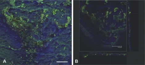
CLSM images of biofilm on soft tissue from patient 2. Overlay projection of the biofilm (A) and a projected side-view (B), meaning the biofilm is visualized from the top and from the side (XZ-plane and YZ-plane, respectively). Bar represents 75 µm. CLSM examination showed that the biofilm distribution was patchy (panel A), with some sites containing large clusters of bacteria and other regions showing hardly any evidence of infection. The bacteria were frequently embedded within a self-produced matrix of EPS (panel B). In addition, the infected ulcer was quite superficial, as can be concluded from the biofilm thickness shown in panel B.
References
-
- Adler AI, Boyko EJ, Ahroni JH, Smith DG. Lower-extremity amputation in diabetes.The independent effects of peripheral vascular disease, sensory neuropathy, and foot ulcers. Diabetes Care. 1999;22:1029–35. - PubMed
-
- Boulton AJ, Vileikyte L, Ragnarson-Tennvall G, Apelqvist J. The global burden of diabetic foot disease. Lancet. 2005;366:1719–24. - PubMed
-
- Costerton JW. Clin Orthop. (437) 2005. Biofilm theory can guide the treatment of device-related orthopedic infections; pp. 7–11. - PubMed
-
- Davis SC, Martinez L, Kirsner R. The diabetic foot: the importance of biofilms and wound bed preparation. Curr Diab Rep. 2006;6:439–45. - PubMed
Publication types
MeSH terms
Substances
LinkOut - more resources
Full Text Sources
Medical