. 2011 May 12;364(19):1795-806.
doi: 10.1056/NEJMoa1101324.

Evidence for human lung stem cells

Affiliations

Evidence for human lung stem cells

Jan Kajstura et al. N Engl J Med. .

Retraction in

Abstract

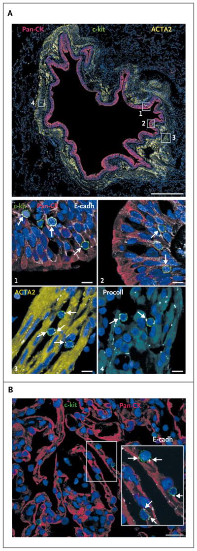
Background: Although progenitor cells have been described in distinct anatomical regions of the lung, description of resident stem cells has remained elusive.

Methods: Surgical lung-tissue specimens were studied in situ to identify and characterize human lung stem cells. We defined their phenotype and functional properties in vitro and in vivo.

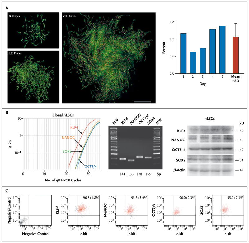
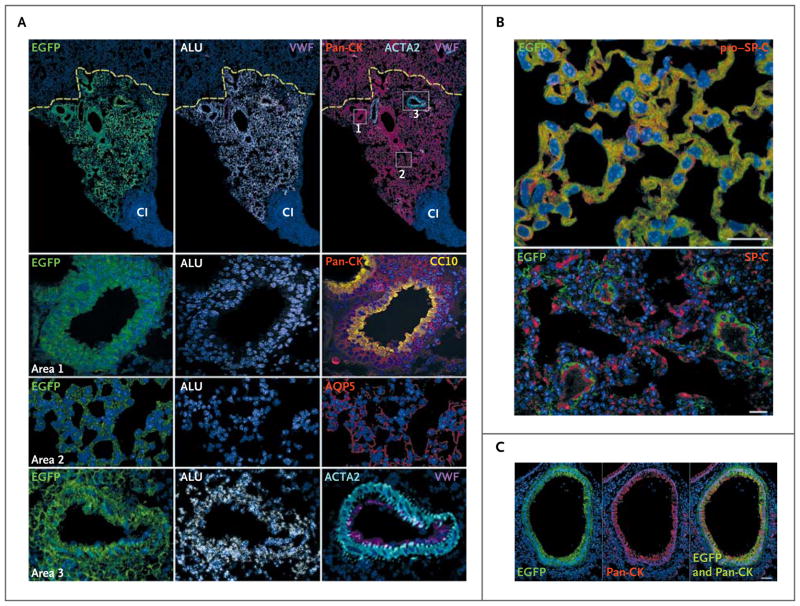
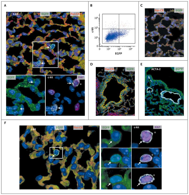
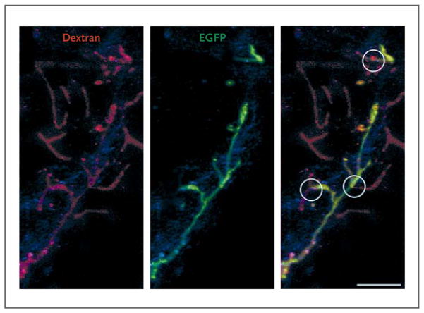
Results: Human lungs contain undifferentiated human lung stem cells nested in niches in the distal airways. These cells are self-renewing, clonogenic, and multipotent in vitro. After injection into damaged mouse lung in vivo, human lung stem cells form human bronchioles, alveoli, and pulmonary vessels integrated structurally and functionally with the damaged organ. The formation of a chimeric lung was confirmed by detection of human transcripts for epithelial and vascular genes. In addition, the self-renewal and long-term proliferation of human lung stem cells was shown in serial-transplantation assays.

Conclusions: Human lungs contain identifiable stem cells. In animal models, these cells participate in tissue homeostasis and regeneration. They have the undemonstrated potential to promote tissue restoration in patients with lung disease. (Funded by the National Institutes of Health.).


Figures

Figure 1
Figure 1. Clonogenicity and Pluripotency of Human Lung Stem Cells (hLSCs)
Panel A shows hLSC clones obtained by limiting dilution. Individual c-kit–positive lineage-negative cells generated clones that increased in size with time. Cloning efficiency is shown in individual cases and as means ±SD. Panel B (left) shows representative results of quantitative reverse-transcriptase–polymerase-chain-reaction (qRT-PCR) assays (with ΔRn indicating the log10-transformed relative number of messenger RNA [mRNA] transcripts) of transcripts for Kruppel-like factor 4 gene (KLF4), the homeobox transcription factor Nanog gene (NANOG), the octamer-binding transcription factor 3–4 gene (OCT3/4), and the sex-determining-region Y–box 2 gene (SOX2) in undifferentiated clonal human lung stem cells. MW denotes molecular-weight DNA ladders. Sequences are listed in Figure 5B in the Supplementary Appendix. The expression of these four genes in undifferentiated clonal hLSCs is also shown (middle, with the transcript sizes shown along the bottom), and the expression of corresponding proteins is also shown (right, with protein sizes listed). β-Actin was used as an indicator of equal loading in all lanes. Panel C shows bivariate-distribution plots (with the x-axis units on a log10 scale and values within each quadrant indicating the mean [±SD] percentage of cells found in each quadrant) of c-kit KLF4, NANOG, OCT3/4, and SOX2 in undifferentiated clonal hLSCs.
Figure 2
Figure 2. Lung Regeneration by Clonal and Nonclonal Human Lung Stem Cells
Regions of cryoinjury (CI) in mouse lung were injected with human lung stem cells, and newly formed human pulmonary structures are visible because the cells express various proteins in various colors (with the protein indicated as the color used in its label in the figure): enhanced green fluorescent protein (EGFP); Alu repeat sequences (ALU); pan-cytokeratin (pan-CK); von Willebrand factor (VWF); Clara cell 10-kD secretory protein (CC10); aquaporin 5 (AQP5); actin, alpha 2, smooth muscle, aorta (ACTA2); and pro–surfactant protein C (pro–SP-C) and SP-C. Lung regeneration was discernible 14 days after delivery of clonal human lung stem cells (Panel A), with the human cells having largely replaced the CI region, adjacent to an area of residual injury. Recipient mouse epithelial cells, positive for pan-CK but negative for EGFP, are present at the periphery of the regenerated tissue; the yellow dashed lines define this boundary (Panel A). Areas within the rectangles are shown at a higher magnification in the lower panels: area 1 corresponds to a human bronchiole composed of epithelial cells positive for EGFP, ALU, and pan-CK and CC10; area 2, human alveoli positive for EGFP, ALU, and AQP5; area 3, a human pulmonary arteriole positive for EGFP, ALU, and ACTA2 and von Willebrand factor (VWF) (Panel A). Panel B shows alveoli, formed by clonal human lung stem cells, that express EGFP and pro–SP-C or EGFP and SP-C (scale bar, 20 μm). Panel C shows newly formed human bronchioles with epithelial cells positive for EGFP and pan-CK (with the merged image shown at the right; scale bar, 50 μm).
Figure 3
Figure 3. Human Lung Stem Cells in the Serial Transplantation Assay
Cells expressing various proteins in various colors (with the protein indicated as the color used in its label in the figure) are shown, as are scatter plots of cells after flow analysis. Panel A shows newly formed human alveoli positive for enhanced green fluorescent protein (EGFP) and pan-cytokeratin (pan-CK) (top). Two c-kit–positive cells are visible (arrows within the rectangle). The c-kit–positive cells are EGFP-positive (bottom left, arrows) and one is cycling, as indicated by its Ki67-positive status (bottom right, asterisk). Panel B shows a scatter plot indicating c-kit–positive, EGFP-positive human lung stem cells (red dots); these were isolated after lung regeneration. After serial transplantation, regenerated alveoli (Panel C; scale bar, 20 μm) and a bronchiole (Panel D; scale bar, 50 μm) are positive for EGFP and pan-CK, whereas a newly formed arteriole (Panel E; scale bar, 50 μm) is positive for EGFP and actin, alpha 2, smooth muscle, aorta (ACTA2). In a lung in which serial transplantation was performed, EGFP-positive, pan-CK–positive human alveoli (Panel F) contain one c-kit–positive cell (arrow within the rectangle). This c-kit–positive cell (and two others, shown individually below the first in the panel on the right) is EGFP-positive (left, arrow) and is cycling (right, asterisk).
Figure 4
Figure 4. Integration of Human Pulmonary Structures
Shown is pulmonary vasculature perfused with rhodamine-labeled dextran. Pulmonary vessels (left, shown in red) that are preexisting have enhanced green fluorescent protein (EGFP)–negative walls (middle), whereas those that are regenerated have EGFP-positive walls (middle). Sites of anastomosis between the preexisting and regenerated pulmonary vessels are shown (circles, right image).
Figure 5
Figure 5. Localization of Human Lung Stem Cells
Cells expressing various proteins in various colors (with the protein indicated as the color used in its label in the figure) are shown. Panel A shows a bronchiole, approximately 900 μm in diameter (scale bar, 0.5 mm in length), with epithelial cells positive for pan-cytokeratin (pan-CK) and smooth-muscle cells expressing actin, alpha 2, smooth muscle, aorta (ACTA2). Several c-kit–positive cells are present within the bronchiolar wall (within the four rectangles); in the images labeled with numerals, these rectangles are shown at higher magnification (scale bar, 10 μm). These show that c-kit–positive cells are connected by E-cadherin (E-cadh, arrows) to bronchiolar epithelial cells (images 1 and 2), smooth-muscle cells (as indicated by ACTA2; image 3, arrows), and fibroblasts (as indicated by procollagen [procoll]; arrows, image 4). The c-kit–positive cells were negative for the markers of mast cells CD45 and tryptase (not shown). Panel B (scale bar, 20 μm) shows that alveoli positive for pan-CK contain c-kit–positive cells in the cell walls. The area within the rectangle is shown at higher magnification in the inset. C-kit–positive cells are connected by E-cadherin (arrows) to epithelial cells.

Comment in

  • Toward lung regeneration.
    Chapman HA. Chapman HA. N Engl J Med. 2011 May 12;364(19):1867-8. doi: 10.1056/NEJMe1101800. N Engl J Med. 2011. PMID: 21561353 No abstract available.
  • Evidence for human lung stem cells.
    Suzuki T, Kondo T, Kubo H. Suzuki T, et al. N Engl J Med. 2011 Aug 4;365(5):465; author reply 465-6. doi: 10.1056/NEJMc1106693. N Engl J Med. 2011. PMID: 21812678 No abstract available.
  • Evidence for human lung stem cells.
    Masuda S. Masuda S. N Engl J Med. 2011 Aug 4;365(5):464-5; author reply 465-6. doi: 10.1056/NEJMc1106693. N Engl J Med. 2011. PMID: 21812679 No abstract available.
  • Evidence for human lung stem cells.
    Moodley Y. Moodley Y. N Engl J Med. 2011 Aug 4;365(5):464; author reply 465-6. doi: 10.1056/NEJMc1106693. N Engl J Med. 2011. PMID: 21812680 No abstract available.
  • Human lung stem cells: oh, the places you'll go!
    Königshoff M, Schwarz J, Eickelberg O. Königshoff M, et al. EMBO Mol Med. 2011 Oct;3(10):575-7. doi: 10.1002/emmm.201100166. Epub 2011 Aug 19. EMBO Mol Med. 2011. PMID: 21853530 Free PMC article. No abstract available.

References

    1. Laugwitz KL, Moretti A, Lam J, et al. Postnatal isl1+ cardioblasts enter fully differentiated cardiomyocyte lineages. Nature. 2005;433:647–53. [Erratum, Nature 2007;446:934.] - PMC - PubMed
    1. Domian IJ, Chiravuri M, van der Meer P, et al. Generation of functional ventricular heart muscle from mouse ventricular progenitor cells. Science. 2009;326:426–9. - PMC - PubMed
    1. Kotton DN, Fine A. Lung stem cells. Cell Tissue Res. 2008;331:145–56. - PubMed
    1. Chen H, Matsumoto K, Stripp BR. Bronchiolar progenitor cells. Proc Am Thorac Soc. 2009;6:602–6. - PMC - PubMed
    1. Rock JR, Onaitis MW, Rawlins EL, et al. Basal cells as stem cells of the mouse trachea and human airway epithelium. Proc Natl Acad Sci U S A. 2009;106:12771–5. - PMC - PubMed

Publication types

Substances