. 2011 Jul 15;286(28):25056-64.
doi: 10.1074/jbc.M110.213777. Epub 2011 May 11.

CCM3/PDCD10 heterodimerizes with germinal center kinase III (GCKIII) proteins using a mechanism analogous to CCM3 homodimerization

Affiliations

CCM3/PDCD10 heterodimerizes with germinal center kinase III (GCKIII) proteins using a mechanism analogous to CCM3 homodimerization

Derek F Ceccarelli et al. J Biol Chem. .

Abstract

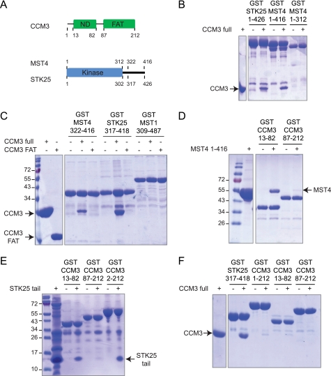
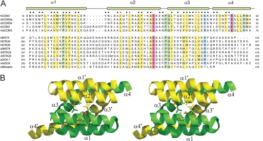
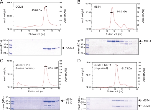
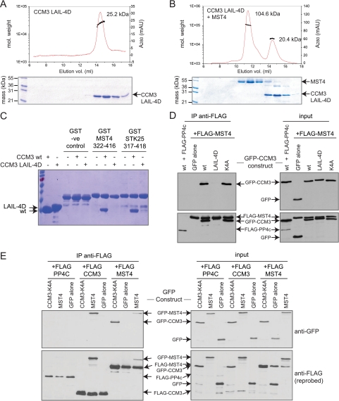
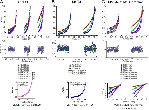
CCM3 mutations give rise to cerebral cavernous malformations (CCMs) of the vasculature through a mechanism that remains unclear. Interaction of CCM3 with the germinal center kinase III (GCKIII) subfamily of Sterile 20 protein kinases, MST4, STK24, and STK25, has been implicated in cardiovascular development in the zebrafish, raising the possibility that dysregulated GCKIII function may contribute to the etiology of CCM disease. Here, we show that the amino-terminal region of CCM3 is necessary and sufficient to bind directly to the C-terminal tail region of GCKIII proteins. This same region of CCM3 was shown previously to mediate homodimerization through the formation of an interdigitated α-helical domain. Sequence conservation and binding studies suggest that CCM3 may preferentially heterodimerize with GCKIII proteins through a manner structurally analogous to that employed for CCM3 homodimerization.


Figures

FIGURE 1.
FIGURE 1.
Purified CCM3 and GCKIII proteins interact directly. A, domain organization of CCM3 and GCKIII proteins. ND, N-terminal dimerization region of CCM3. B, in vitro interaction between GST fusions of STK25 and MST4 with CCM3. The minimal MST4 kinase domain does not bind CCM3. C, CCM3 interacts with the C-terminal tail regions of MST4 and STK25 but not MST1. The CCM3 C-terminal FAT domain does not interact with the kinases. D, the amino terminus of CCM3 (CCM3-ND) is sufficient for interaction with full length MST4. E, CCM3-ND is sufficient for interaction with the C-terminal tail region of STK25. F, CCM3 binds tightly to the GST-STK25 tail but not to GST-CCM3.
FIGURE 2.
FIGURE 2.
Sequence conservation between CCM3 and GCKIII proteins. A, sequence alignment of CCM3 N-terminal and GCKIII protein C-terminal tails regions. h, human; dr, Danio rerio; e, C. elegans; mb, Monosiga brevicollis; and dd, Dictyostelium discoidium sequences of CCM3 and GCKIII proteins (STK24, MST4, STK25, and severin) are shown. Conserved hydrophobic, acidic, basic, and proline/glycine residues are highlighted in yellow, red, blue, and green, respectively. Residues comprising the CCM3 homodimer interface are indicated by circles (●) at the top of the alignment. The CCM3 residues (Leu-44, Ala047, Ile-66, and Leu-67) mutated in this study are highlighted with red circles. B, schematic of the N-terminal dimerization region of CCM3 (Protein Data Bank code 3L8I). Protomer chains are colored yellow and green with residues mutated in this study shown in stick representation. Secondary structure elements are labeled.
FIGURE 3.
FIGURE 3.
Size exclusion chromatography and multiangle light scattering of purified CCM3 and MST4 proteins. The left vertical axis denotes molecular weight of eluting species. The right vertical axis denotes absorbance measurement of eluent. The area of the peak integrated for analysis of molecular mass is indicated by a black line. An SDS-PAGE analysis of the corresponding eluted proteins is shown below each chromatogram. A, CCM3 alone. B, MST4 full-length alone (the asterisk indicates a likely MST4 degradation product). C, MST4 kinase domain (residues 1–312). D, co-purified CCM3-MST4 complex.
FIGURE 4.
FIGURE 4.
Mutations in the amino terminus of CCM3 disrupt the CCM3-GCKIII heterodimer interaction in vitro and in vivo. A, the N-terminal CCM3 mutant (LAIL-4D) elutes as a monomer, as assessed by SEC-MALS. mAU, milli absorbance units. B, the N-terminal CCM3 mutant (LAIL-4D) does not co-elute with full-length wild type MST4, as assessed by SEC-MALS. C, the N-terminal CCM3 mutant (LAIL-4D) does not interact with MST4 or STK25 tail regions in a GST pulldown assay. D, FLAG-MST4 interaction with GFP-CCM3 in transiently transfected HEK293T cells is disrupted by mutations in the N-terminal region of CCM3 (LAIL-4D) but not by mutations within the FAT domain (K4A) of CCM3. FLAG-tagged PP4C was used as a negative binding control. Right panels show expression in the cell lysate; left panels show immunoprecipitated proteins. Top panels are blotted with anti-GFP antibody; bottom panels have been reprobed with anti-FLAG. E, FLAG-CCM3 and FLAG-MST4 interact strongly with GFP-MST4 and GFP-CCM3, respectively, in HEK293T cells. A weaker interaction of GFP-MST4 with FLAG-MST4 was detected. (Note that this interaction may be mediated via dimerization of other STRIPAK components.) No interaction of FLAG-CCM3 with GFP-CCM3 was detected. To eliminate detection of indirect interactions between GFP-CCM3 and FLAG-CCM3 arising from bridging interactions with a dimeric STRIPAK complex, we employed a four site mutant within the FAT domain of CCM3 (in the context of the GFP-CCM3 construct) that abolishes interaction with STRIPAK. FLAG-tagged PP4C was used as a negative binding control. Right panels show expression in the cell lysate; left panels show immunoprecipitated proteins. Top panels are blotted with anti-GFP antibody; bottom panels have been reprobed with anti-FLAG. vol., volume.
FIGURE 5.
FIGURE 5.
Analytical ultracentrifugation analysis of CCM3 and MST4 dimerization potential. A, sedimentation equilibrium analysis of CCM3. Sedimentation equilibrium ultracentrifugation was performed at 25 °C and spin speeds of 10,000, 15,000, 20,000, and 25,000 rpm. Global fits to a dimer-monomer equilibrium model performed on 12 data sets (top panel), which were acquired at four spin speeds and three spin radii. This model yielded a good fit and a measured Kd value of 2.7 ± 0.8 μm. Residuals to the monomer-dimer equilibrium fits are shown in the middle panel. Fit to a single-species dimer model (lower panel) yielded poor agreement. B, sedimentation equilibrium analysis of MST4. Sedimentation equilibrium ultracentrifugation was performed at 25 °C and spin speeds of 10,000, 15,000, and 20,000 rpm. Global fits to a dimer-monomer equilibrium model performed on six data sets (top panel), which were acquired at three spin speeds and two spin radii. This model yielded a good fit and a measured KD value of 2.5 ± 0.4 μm. Residuals to the monomer-dimer equilibrium fits are shown in middle panel. Fit to a single-species dimer model (lower panel) yielded poor agreement. C, sedimentation equilibrium analysis of an equimolar CCM3-MST4 complex. Sedimentation equilibrium ultracentrifugation was performed at 25 °C and spin speeds of 10,000, 15,000, 20,000, and 25,000 rpm. Global fits to a single-species model performed on eight data sets (top panel) acquired at four spin speeds and two spin radii. With the mass of the heterodimer input as an initial parameter, this model yielded a good fit. Residuals to the monomer-dimer equilibrium fits are shown in middle panel. Plot of the natural logarithm of the A280 value versus the square of the spin radius (lower panel). All spin speeds yield straight lines, consistent with only one species present at detectable levels at equilibrium.

References

    1. Labauge P., Denier C., Bergametti F., Tournier-Lasserve E. (2007) Lancet Neurol. 6, 237–244 - PubMed
    1. Revencu N., Vikkula M. (2006) J. Med. Genet. 43, 716–721 - PMC - PubMed
    1. Bergametti F., Denier C., Labauge P., Arnoult M., Boetto S., Clanet M., Coubes P., Echenne B., Ibrahim R., Irthum B., Jacquet G., Lonjon M., Moreau J. J., Neau J. P., Parker F., Tremoulet M., Tournier-Lasserve E. (2005) Am. J. Hum. Genet. 76, 42–51 - PMC - PubMed
    1. Guclu B., Ozturk A. K., Pricola K. L., Bilguvar K., Shin D., O'Roak B. J., Gunel M. (2005) Neurosurgery 57, 1008–1013 - PubMed
    1. Zheng X., Xu C., Di Lorenzo A., Kleaveland B., Zou Z., Seiler C., Chen M., Cheng L., Xiao J., He J., Pack M. A., Sessa W. C., Kahn M. L. (2010) J. Clin. Invest. 120, 2795–2804 - PMC - PubMed

Publication types

MeSH terms