. 2011 Jul;85(14):7203-15.
doi: 10.1128/JVI.00262-11. Epub 2011 May 11.

Herpes simplex virus 1 pUL34 plays a critical role in cell-to-cell spread of virus in addition to its role in virus replication

Affiliations

Herpes simplex virus 1 pUL34 plays a critical role in cell-to-cell spread of virus in addition to its role in virus replication

Alison C Haugo et al. J Virol. 2011 Jul.

Abstract

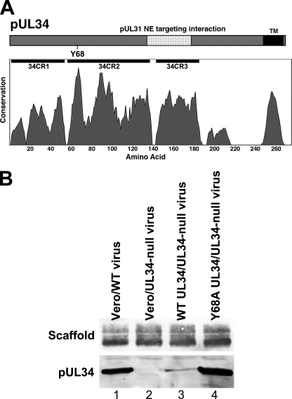
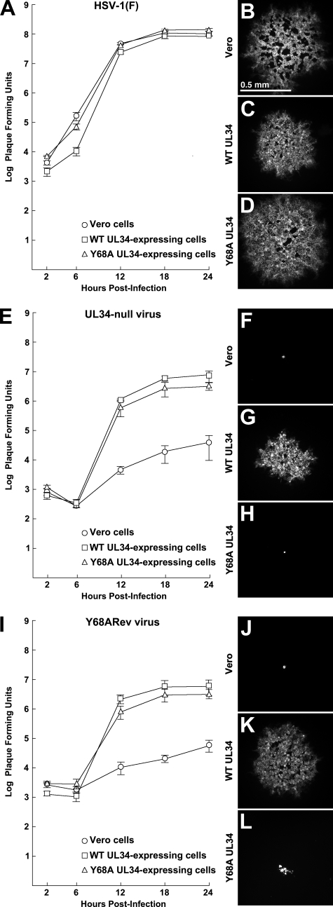
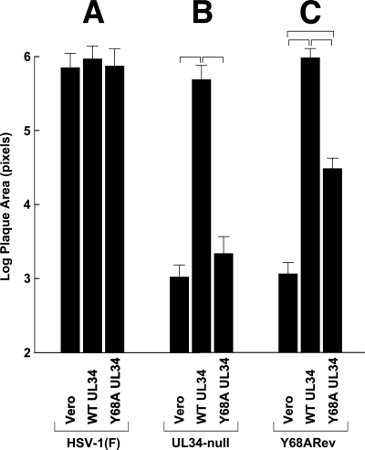
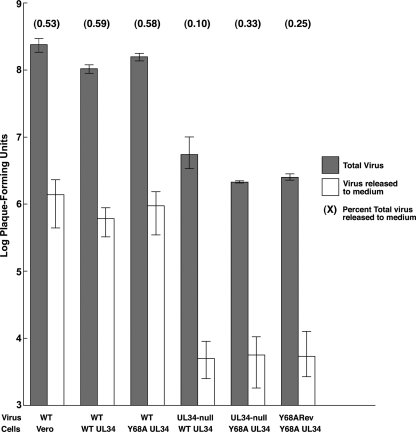
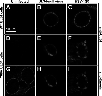
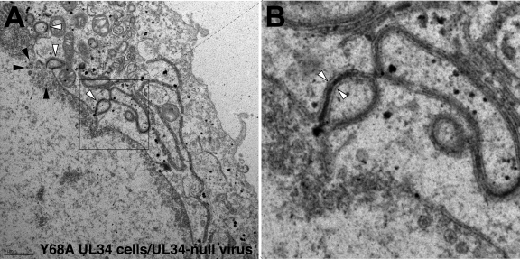
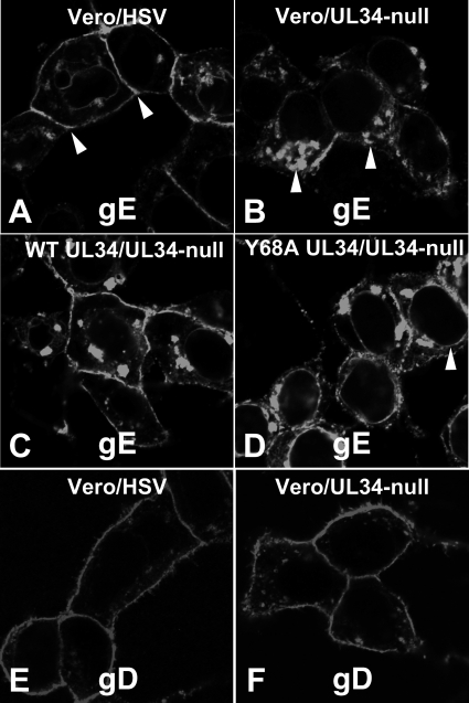
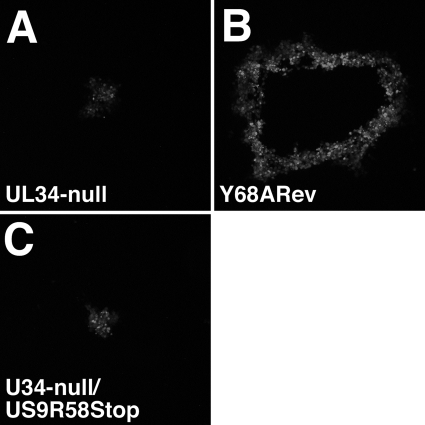
Herpes simplex virus (HSV) pUL34 plays a critical role in virus replication by mediating egress of nucleocapsids from the infected cell nucleus. We have identified a mutation in pUL34 (Y68A) that produces a major defect in virus replication and impaired nuclear egress but also profoundly inhibits cell-to-cell spread and trafficking of gE. Virion release to the extracellular medium is not affected by the Y68A mutation, indicating that the mutation specifically inhibits cell-to-cell spread. We isolated extragenic suppressors of the Y68A plaque formation defect and mapped them by a combination of high-throughput Illumina sequencing and PCR-based screening. We found that suppression is highly correlated with a nonsense mutation in the US9 gene, which plays a critical role in cell-to-cell spread of HSV-1 in neurons. The US9 mutation alone is not sufficient to suppress the Y68A spread phenotype, indicating a likely role for multiple viral factors.


Figures

Fig. 1.
Fig. 1.
Position of the Y68A mutation and expression of pUL34 by cell lines. (A) Schematic diagrams of pUL34 showing the locations of relevant sequence features. Protein sequence of pUL34 is indicated as a bar with the N terminus at the left. Sequences in pUL34 that mediate nuclear envelope targeting of the NEC are indicated as a stippled region. Position of Y68 is indicated below the bar. Positions of conserved regions are indicated immediately below each of the bars. The conservation plot shows conservation of biochemical properties of amino acids using all available herpesvirus sequences aligned by using the program MUSCLE (13). (B) Expression of wild-type and Y68A mutant pUL34 by stable cell lines. Digital images of Western blots are shown. Vero cells (lanes 1 and 2) or cells stably expressing WT pUL34 (lane 3) or Y68A pUL34 (lane 4) were infected with WT HSV-1(F) (lane 1) or UL34-null vRR1072(TK+) virus (lanes 2 to 4). Blotted infected cell proteins were probed for either scaffolding protein (top) or pUL34 (bottom).
Fig. 2.
Fig. 2.
Single-step growth and plaque formation on Vero, WT pUL34-expressing, or Y68A mutant pUL34-expressing cell lines. For single-step growth replicate, cultures of Vero cells, WT UL34-express-ing cells, or Y68A UL34-expressing cells were infected at an MOI of 5 with HSV-1(F) (A), the UL34-null virus vRR1072(TK+) (E), or Y68ARev (I). Residual virus was removed or inactivated with a low-pH wash, and at the indicated times total culture virus was titrated on WT UL34-expressing cells. Virus yields are expressed as PFU per milliliter. Each data point represents the mean of results from three independent experiments. Error bars indicate the range of values. For plaque formation assays, digital micrographs of infected cell monolayers stained for glycoprotein D are shown. The cell line infected is indicated to the left of each panel. In panels B to D, the infecting virus was HSV-1(F). In panels F to H, the infecting virus was the UL34-null virus vRR1072(TK+). In panels J to L, the infecting virus was Y68ARev. All plaques were fixed and stained at 2 days after infection. All plaque images are shown at the same magnification.
Fig. 3.
Fig. 3.
Quantitation of plaque formation on wild-type and mutant UL34-expressing cell lines. Histograms of mean plaque sizes on Vero, WT pUL34-expressing, and Y68A pUL34-expressing cells are shown. Plaques stained 2 days after infection, as described in the legend to Fig. 2, were photographed, and plaque areas in image pixels were determined using ImageJ. For each bar, 20 randomly selected plaques from two independent experiments (40 plaques total) were measured. Brackets indicate pairwise statistical comparisons performed using a Student t test. All of the indicated comparisons showed a highly significant difference (P < 0.001). Note that the y axis has a logarithmic scale.
Fig. 4.
Fig. 4.
Virus release to the medium mediated by wild-type and mutant pUL34. Replicate cultures of Vero, wild-type pUL34-expressing, or Y68A mutant pUL34-expressing cells were infected at an MOI of 5 with wild-type virus HSV-1(F), UL34-null virus vRR1072(TK+), or Y68ARev. Residual virus was removed or inactivated with a low-pH wash, and at 18 h after infection total culture virus (gray bars) or culture supernatant (white bars) was titrated on WT UL34-expressing cells. Virus yields are expressed as PFU/ml. Each data point represents the mean of results from three independent experiments. Error bars indicate the range of values. The values in parentheses above each pair of bars indicate the percentages of total virus released to the medium calculated as the mean amount of released virus divided by the mean amount of total virus times 100. Low pH wash completely removed residual extracellular virus, as the titer of culture supernatant at 2 h postinfection was undetectable.
Fig. 5.
Fig. 5.
Localization of WT pUL34, Y68A mutant pUL34, and emerin in infected cells. Digital confocal images of WT UL34-expressing cells (A to C) or Y68A UL34-expressing cells (D to I) are shown. Cells were mock infected (A, D, and G) or infected for 16 h with UL34-null virus (B, E, and H) or with HSV-1(F) (C, F, and I) and then fixed and immunofluorescently stained for pUL34 (A to F) or emerin (G to I). All images are shown at the same magnification.
Fig. 6.
Fig. 6.
TEM analysis of cells that express Y68A UL34. Digital micrographs show Y68A UL34-expressing cells infected with the UL34-null virus vRR1072(TK+) for 20 h. White arrowheads in panel A point to examples of blebbing of the nuclear membrane into the cytoplasm. Black arrowheads point to examples of C capsids in the nucleus. The boxed area in panel A is enlarged (×4) in panel B. The white arrowheads in panel B point to an instance of a bleb with two thicknesses of nuclear envelope separated by an electron-dense layer.
Fig. 7.
Fig. 7.
Wild-type pUL34 expression is required for normal localization of gE but not gD. Vero cells (A, B, E, and F), WT UL34-expressing cells (C), and Y68A UL34-expressing cells (D) were infected with HSV-1(F) (A and E) or UL34-null virus (B to D and F) for 16 h and then fixed and immunofluorescently stained for gE (A to D) or gD (E and F). Arrowheads in panel A point to junctional surfaces, where gE concentrates in wild-type infection. Arrowheads in panel B point to cytoplasmic membranes, where gE concentrates during UL34-null infection. Arrowhead in panel D points to the nuclear envelope.
Fig. 8.
Fig. 8.
Characterization of mutation at the UL34 and US9 loci. Digital images show electrophoretically separated PCR products. (A) Amplification products from the UL34 locus using genomes from rescued viruses as the template. The BAC used for rescue is indicated above each lane. The sizes of the PCR products are indicated to the right of the gel. Lambda BstEII digest size standards are shown in lane 1, and the sizes of standard bands are indicated to the left of the gel. (B) Amplification products from the US9 locus that are either undigested (lanes 2 and 4) or digested with restriction enzymes HincII (lanes 3 and 5). A 100-bp ladder is shown in lane 1.
Fig. 9.
Fig. 9.
Growth of BAC-derived UL34-null and US9R58Stop virus on WT and mutant pUL34-expressing cells. Digital micrographs are shown of representative plaques formed for 1 week on Y68A pUL34-expressing cells. Plaques were immunofluorescently stained for gD. The infecting virus is indicated in each panel.

Similar articles

Cited by

References

    1. Balan P., et al. 1994. An analysis of the in vitro and in vivo phenotypes of mutants of herpes simplex virus type 1 lacking glycoproteins gG, gE, gI or the putative gJ. J. Gen. Virol. 75:1245–1258 - PubMed
    1. Bjerke S. L., et al. 2003. Effects of charged cluster mutations on the function of herpes simplex virus type 1 UL34 protein. J. Virol. 77:7601–7610 - PMC - PubMed
    1. Bjerke S. L., Roller R. 2006. Roles for herpes simplex type 1 UL34 and US3 proteins in disrupting the nuclear lamina during herpes simplex virus type 1 egress. Virology 347(2):261–276 - PMC - PubMed
    1. Brideau A. D., Banfield B. W., Enquist L. W. 1998. The Us9 gene product of pseudorabies virus, an alphaherpesvirus, is a phosphorylated, tail-anchored type II membrane protein. J. Virol. 72:4560–4570 - PMC - PubMed
    1. Brideau A. D., Card J. P., Enquist L. W. 2000. Role of pseudorabies virus Us9, a type II membrane protein, in infection of tissue culture cells and the rat nervous system. J. Virol. 74:834–845 - PMC - PubMed

Publication types